 Movie
Movie Controller

[English] 日本語
 Yorodumi
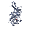
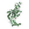
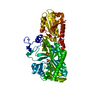
- PDB-7kis: Crystal structure of Pseudomonas aeruginosa PBP2 in complex with ... -
+ Open data

- Basic information
| Entry | Database: PDB / ID: 7kis | ||||||
|---|---|---|---|---|---|---|---|
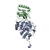
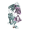
| Title | Crystal structure of Pseudomonas aeruginosa PBP2 in complex with WCK 5153 | ||||||
 Components Components | Peptidoglycan D,D-transpeptidase MrdA | ||||||
 Keywords Keywords | HYDROLASE/Inhibitor/Antibiotic / Inhibitor / cell wall / antibiotic resistance / HYDROLASE / HYDROLASE-Inhibitor-Antibiotic complex | ||||||
| Function / homology |  Function and homology information Function and homology informationserine-type D-Ala-D-Ala carboxypeptidase / serine-type D-Ala-D-Ala carboxypeptidase activity / penicillin binding / peptidoglycan biosynthetic process / cell wall organization / regulation of cell shape / proteolysis / plasma membrane Similarity search - Function | ||||||
| Biological species |   Pseudomonas aeruginosa (bacteria) Pseudomonas aeruginosa (bacteria) | ||||||
| Method |  X-RAY DIFFRACTION / SYNCHROTRON / MOLECULAR REPLACEMENT / Resolution: 2.869 Å X-RAY DIFFRACTION / SYNCHROTRON / MOLECULAR REPLACEMENT / Resolution: 2.869 Å | ||||||
 Authors Authors | Rajavel, M. / van den Akker, F. | ||||||
 Citation Citation | Journal: Mbio / Year: 2021 Title: Structural Characterization of Diazabicyclooctane beta-Lactam "Enhancers" in Complex with Penicillin-Binding Proteins PBP2 and PBP3 of Pseudomonas aeruginosa. Authors: Rajavel, M. / Kumar, V. / Nguyen, H. / Wyatt, J. / Marshall, S.H. / Papp-Wallace, K.M. / Deshpande, P. / Bhavsar, S. / Yeole, R. / Bhagwat, S. / Patel, M. / Bonomo, R.A. / van den Akker, F. | ||||||
| History |
|
- Structure visualization
| Structure viewer | Molecule: MolmilJmol/JSmol |
|---|
- Downloads & links
-Download
| PDBx/mmCIF format | 7kis.cif.gz | 191.7 KB | Display | PDBx/mmCIF format |
|---|---|---|---|---|
| PDB format | pdb7kis.ent.gz | 148.7 KB | Display | PDB format |
| PDBx/mmJSON format | 7kis.json.gz | Tree view | PDBx/mmJSON format | |
| Others |  Other downloads Other downloads |
-Validation report
| Arichive directory | https://data.pdbj.org/pub/pdb/validation_reports/ki/7kisftp://data.pdbj.org/pub/pdb/validation_reports/ki/7kis | HTTPS FTP |
|---|
-Related structure data
| Related structure data | 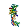 7kitC 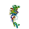 7kivC 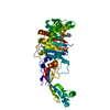 7kiwC 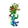 6g9fS S: Starting model for refinement C: citing same article ( |
|---|---|
| Similar structure data |


-Links
 PDBj
PDBj
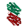
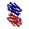
- Assembly
| Deposited unit | 
| |||||||||||||||||||||||||||||||||||||||||||||||||||||||||||||||||||
|---|---|---|---|---|---|---|---|---|---|---|---|---|---|---|---|---|---|---|---|---|---|---|---|---|---|---|---|---|---|---|---|---|---|---|---|---|---|---|---|---|---|---|---|---|---|---|---|---|---|---|---|---|---|---|---|---|---|---|---|---|---|---|---|---|---|---|---|---|
| 1 | 
| |||||||||||||||||||||||||||||||||||||||||||||||||||||||||||||||||||
| 2 | 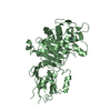
| |||||||||||||||||||||||||||||||||||||||||||||||||||||||||||||||||||
| Unit cell |
| |||||||||||||||||||||||||||||||||||||||||||||||||||||||||||||||||||
| Noncrystallographic symmetry (NCS) | NCS domain:
NCS domain segments:
|