| 登録情報 | データベース: PDB / ID: 6qk1
|
|---|
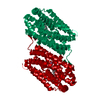
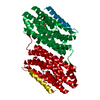
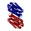
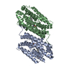
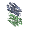
| タイトル | R2-like ligand-binding oxidase Y175F mutant with aerobically reconstituted Mn/Fe cofactor |
|---|
 要素 要素 | Ribonucleotide reductase small subunit |
|---|
 キーワード キーワード | OXIDOREDUCTASE / R2-LIKE LIGAND-BINDING OXIDASE / MN/FE COFACTOR / RIBONUCLEOTIDE REDUCTASE R2 SUBUNIT FOLD / METALLOPROTEIN OXIDOREDUCTASE |
|---|
| 機能・相同性 |  機能・相同性情報 機能・相同性情報
deoxyribonucleotide biosynthetic process / oxidoreductase activity / metal ion binding類似検索 - 分子機能 R2-like ligand binding oxidase / Ribonucleotide Reductase, subunit A / Ribonucleotide Reductase, subunit A / Ribonucleotide reductase small subunit family / Ribonucleotide reductase, small chain / Ribonucleotide reductase-like / Ferritin-like superfamily / Orthogonal Bundle / Mainly Alpha類似検索 - ドメイン・相同性 : / : / MANGANESE (III) ION / PALMITIC ACID / R2-like ligand binding oxidase類似検索 - 構成要素 |
|---|
| 生物種 |  Geobacillus kaustophilus HTA426 (バクテリア) Geobacillus kaustophilus HTA426 (バクテリア) |
|---|
| 手法 |  X線回折 / シンクロトロン / フーリエ合成 / 解像度: 1.754 Å X線回折 / シンクロトロン / フーリエ合成 / 解像度: 1.754 Å |
|---|
 データ登録者 データ登録者 | Griese, J.J. / Hogbom, M. |
|---|
 引用 引用 | ジャーナル: J.Am.Chem.Soc. / 年: 2020
タイトル: Key Structural Motifs Balance Metal Binding and Oxidative Reactivity in a Heterobimetallic Mn/Fe Protein.
著者: Kisgeropoulos, E.C. / Griese, J.J. / Smith, Z.R. / Branca, R.M.M. / Schneider, C.R. / Hogbom, M. / Shafaat, H.S. |
|---|
| 履歴 | | 登録 | 2019年1月28日 | 登録サイト: PDBE / 処理サイト: PDBE |
|---|
| 改定 1.0 | 2020年2月19日 | Provider: repository / タイプ: Initial release |
|---|
| 改定 1.1 | 2020年8月26日 | Group: Database references / Derived calculations / カテゴリ: citation / citation_author / struct_conn
Item: _citation.country / _citation.journal_abbrev ..._citation.country / _citation.journal_abbrev / _citation.journal_id_ASTM / _citation.journal_id_CSD / _citation.journal_id_ISSN / _citation.journal_volume / _citation.page_first / _citation.page_last / _citation.pdbx_database_id_DOI / _citation.pdbx_database_id_PubMed / _citation.title / _citation.year / _struct_conn.pdbx_dist_value / _struct_conn.ptnr1_label_atom_id / _struct_conn.ptnr2_auth_comp_id / _struct_conn.ptnr2_auth_seq_id / _struct_conn.ptnr2_label_asym_id / _struct_conn.ptnr2_label_atom_id / _struct_conn.ptnr2_label_comp_id |
|---|
| 改定 1.2 | 2024年1月24日 | Group: Data collection / Database references / Refinement description
カテゴリ: chem_comp_atom / chem_comp_bond ...chem_comp_atom / chem_comp_bond / database_2 / pdbx_initial_refinement_model
Item: _database_2.pdbx_DOI / _database_2.pdbx_database_accession |
|---|
|
|---|
 ムービー
ムービー コントローラー


 データを開く
 基本情報
基本情報 構造の表示
構造の表示 ダウンロードとリンク
 その他のダウンロード


 PDBj
PDBj


 集合体
集合体


 分子量: 256.424 Da / 分子数: 1 / 由来タイプ: 合成 / 式: C16H32O2
 分子量: 54.938 Da / 分子数: 1 / 由来タイプ: 合成 / 式: Mn
 分子量: 54.938 Da / 分子数: 1 / 由来タイプ: 合成 / 式: Mn
 分子量: 55.845 Da / 分子数: 1 / 由来タイプ: 合成 / 式: Fe
 試料調製
試料調製 / ビームライン: X06SA / 波長: 0.9999 Å
 解析
解析