 Movie
Movie Controller

[English] 日本語
 Yorodumi
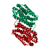
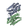
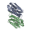
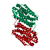
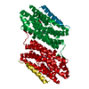
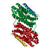
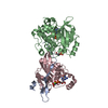
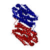
- PDB-6qjv: R2-like ligand-binding oxidase E69D mutant with aerobically recon... -
+ Open data

- Basic information
| Entry | Database: PDB / ID: 6qjv | ||||||
|---|---|---|---|---|---|---|---|
| Title | R2-like ligand-binding oxidase E69D mutant with aerobically reconstituted Mn/Fe cofactor | ||||||
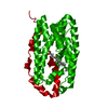
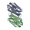
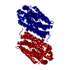
 Components Components | Ribonucleotide reductase small subunit | ||||||
 Keywords Keywords | OXIDOREDUCTASE / R2-LIKE LIGAND-BINDING OXIDASE / MN/FE COFACTOR / RIBONUCLEOTIDE REDUCTASE R2 SUBUNIT FOLD / METALLOPROTEIN OXIDOREDUCTASE | ||||||
| Function / homology |  Function and homology information Function and homology informationdeoxyribonucleotide biosynthetic process / oxidoreductase activity / metal ion binding Similarity search - Function | ||||||
| Biological species |  Geobacillus kaustophilus (bacteria) Geobacillus kaustophilus (bacteria) | ||||||
| Method |  X-RAY DIFFRACTION / SYNCHROTRON / FOURIER SYNTHESIS / Resolution: 1.905 Å X-RAY DIFFRACTION / SYNCHROTRON / FOURIER SYNTHESIS / Resolution: 1.905 Å | ||||||
 Authors Authors | Griese, J.J. / Hogbom, M. | ||||||
 Citation Citation | Journal: J.Am.Chem.Soc. / Year: 2020 Title: Key Structural Motifs Balance Metal Binding and Oxidative Reactivity in a Heterobimetallic Mn/Fe Protein. Authors: Kisgeropoulos, E.C. / Griese, J.J. / Smith, Z.R. / Branca, R.M.M. / Schneider, C.R. / Hogbom, M. / Shafaat, H.S. | ||||||
| History |
|
- Structure visualization
| Structure viewer | Molecule: MolmilJmol/JSmol |
|---|
- Downloads & links
-Download
| PDBx/mmCIF format | 6qjv.cif.gz | 126.8 KB | Display | PDBx/mmCIF format |
|---|---|---|---|---|
| PDB format | pdb6qjv.ent.gz | 98.4 KB | Display | PDB format |
| PDBx/mmJSON format | 6qjv.json.gz | Tree view | PDBx/mmJSON format | |
| Others |  Other downloads Other downloads |
-Validation report
| Arichive directory | https://data.pdbj.org/pub/pdb/validation_reports/qj/6qjvftp://data.pdbj.org/pub/pdb/validation_reports/qj/6qjv | HTTPS FTP |
|---|
-Related structure data
| Related structure data |  6qk0C  6qk1C  6qk2C 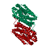 4hr0S S: Starting model for refinement C: citing same article ( |
|---|---|
| Similar structure data |


-Links
 PDBj
PDBj


- Assembly
| Deposited unit | 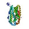
| |||||||||
|---|---|---|---|---|---|---|---|---|---|---|
| 1 | 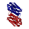
| |||||||||
| Unit cell |
| |||||||||
| Components on special symmetry positions |
|
-Components
-Protein , 1 types, 1 molecules A
| #1: Protein | Mass: 36948.781 Da / Num. of mol.: 1 / Mutation: E69D Source method: isolated from a genetically manipulated source Source: (gene. exp.) Geobacillus kaustophilus (strain HTA426) (bacteria)Strain: HTA426 / Gene: GK2771 / Plasmid: pET-46 Ek/LIC / Production host: References: UniProt: Q5KW80, ribonucleoside-diphosphate reductase |
|---|
-Non-polymers , 5 types, 116 molecules 


| #2: Chemical | ChemComp-PLM /  Mass: 256.424 Da / Num. of mol.: 1 / Source method: obtained synthetically / Formula: C16H32O2 Mass: 256.424 Da / Num. of mol.: 1 / Source method: obtained synthetically / Formula: C16H32O2 |
|---|---|
| #3: Chemical | ChemComp-MN3 /  Mass: 54.938 Da / Num. of mol.: 1 / Source method: obtained synthetically / Formula: Mn Mass: 54.938 Da / Num. of mol.: 1 / Source method: obtained synthetically / Formula: Mn |
| #4: Chemical | ChemComp-MN /  Mass: 54.938 Da / Num. of mol.: 1 / Source method: obtained synthetically / Formula: Mn Mass: 54.938 Da / Num. of mol.: 1 / Source method: obtained synthetically / Formula: Mn |
| #5: Chemical | ChemComp-FE /  Mass: 55.845 Da / Num. of mol.: 1 / Source method: obtained synthetically / Formula: Fe Mass: 55.845 Da / Num. of mol.: 1 / Source method: obtained synthetically / Formula: Fe |
| #6: Water | ChemComp-HOH / Mass: 18.015 Da / Num. of mol.: 112 / Source method: isolated from a natural source / Formula: H2O |
-Experimental details
-Experiment
| Experiment | Method: X-RAY DIFFRACTION / Number of used crystals: 1 |
|---|
- Sample preparation
| Crystal | Density Matthews: 2.36 Å3/Da / Density % sol: 47.98 % |
|---|---|
| Crystal grow | Temperature: 295 K / Method: vapor diffusion, hanging drop / pH: 7.4 / Details: 30% (W/V) PEG 1500, 0.1 M HEPES-NA PH 7.4 |
-Data collection
| Diffraction | Mean temperature: 100 K / Serial crystal experiment: N |
|---|---|
| Diffraction source | Source: SYNCHROTRON / Site: SLS  / Beamline: X06SA / Wavelength: 0.9999 Å / Beamline: X06SA / Wavelength: 0.9999 Å |
| Detector | Type: DECTRIS PILATUS 6M / Detector: PIXEL / Date: Nov 28, 2012 |
| Radiation | Protocol: SINGLE WAVELENGTH / Monochromatic (M) / Laue (L): M / Scattering type: x-ray |
| Radiation wavelength | Wavelength: 0.9999 Å / Relative weight: 1 |
| Reflection | Resolution: 1.9→50 Å / Num. obs: 27535 / % possible obs: 98.8 % / Observed criterion σ(I): -3 / Redundancy: 3.3 % / Biso Wilson estimate: 30.62 Å2 / CC1/2: 0.999 / Rmerge(I) obs: 0.05 / Rrim(I) all: 0.06 / Net I/σ(I): 14.49 |
| Reflection shell | Resolution: 1.9→2.02 Å / Redundancy: 3.2 % / Rmerge(I) obs: 0.658 / Mean I/σ(I) obs: 1.79 / Num. unique obs: 4310 / CC1/2: 0.691 / Rrim(I) all: 0.79 / % possible all: 97.1 |
- Processing
| Software |
| |||||||||||||||||||||||||||||||||||||||||||||||||||||||||||||||||||||||||||||
|---|---|---|---|---|---|---|---|---|---|---|---|---|---|---|---|---|---|---|---|---|---|---|---|---|---|---|---|---|---|---|---|---|---|---|---|---|---|---|---|---|---|---|---|---|---|---|---|---|---|---|---|---|---|---|---|---|---|---|---|---|---|---|---|---|---|---|---|---|---|---|---|---|---|---|---|---|---|---|
| Refinement | Method to determine structure: FOURIER SYNTHESIS Starting model: 4hr0 Resolution: 1.905→39.35 Å / SU ML: 0.22 / Cross valid method: FREE R-VALUE / σ(F): 2 / Phase error: 20.89
| |||||||||||||||||||||||||||||||||||||||||||||||||||||||||||||||||||||||||||||
| Solvent computation | Shrinkage radii: 0.9 Å / VDW probe radii: 1.11 Å | |||||||||||||||||||||||||||||||||||||||||||||||||||||||||||||||||||||||||||||
| Refinement step | Cycle: LAST / Resolution: 1.905→39.35 Å
| |||||||||||||||||||||||||||||||||||||||||||||||||||||||||||||||||||||||||||||
| Refine LS restraints |
| |||||||||||||||||||||||||||||||||||||||||||||||||||||||||||||||||||||||||||||
| LS refinement shell |
|
