 Movie
Movie Controller

[English] 日本語
 Yorodumi
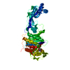
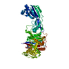
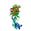
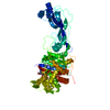
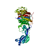
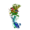
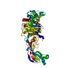
- PDB-6vot: Crystal structure of Pseudomonas aerugonisa PBP3 complexed to gam... -
+ Open data

- Basic information
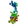
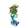
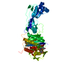
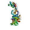
| Entry | Database: PDB / ID: 6vot | ||||||
|---|---|---|---|---|---|---|---|
| Title | Crystal structure of Pseudomonas aerugonisa PBP3 complexed to gamma-lactam YU253434 | ||||||
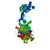
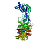
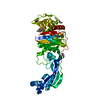
 Components Components | Peptidoglycan D,D-transpeptidase FtsI | ||||||
 Keywords Keywords | HYDROLASE / Inhibitor complex / penicillin-binding protein / ANTIBIOTIC | ||||||
| Function / homology |  Function and homology information Function and homology informationpeptidoglycan glycosyltransferase activity / serine-type D-Ala-D-Ala carboxypeptidase / serine-type D-Ala-D-Ala carboxypeptidase activity / division septum assembly / FtsZ-dependent cytokinesis / penicillin binding / peptidoglycan biosynthetic process / cell wall organization / regulation of cell shape / proteolysis / plasma membrane Similarity search - Function | ||||||
| Biological species |   Pseudomonas aeruginosa (bacteria) Pseudomonas aeruginosa (bacteria) | ||||||
| Method |  X-RAY DIFFRACTION / SYNCHROTRON / MOLECULAR REPLACEMENT / Resolution: 2.4 Å X-RAY DIFFRACTION / SYNCHROTRON / MOLECULAR REPLACEMENT / Resolution: 2.4 Å | ||||||
 Authors Authors | van den Akker, F. | ||||||
 Citation Citation | Journal: J.Med.Chem. / Year: 2020 Title: A gamma-Lactam Siderophore Antibiotic Effective against Multidrug-Resistant Gram-Negative Bacilli. Authors: Goldberg, J.A. / Nguyen, H. / Kumar, V. / Spencer, E.J. / Hoyer, D. / Marshall, E.K. / Cmolik, A. / O'Shea, M. / Marshall, S.H. / Hujer, A.M. / Hujer, K.M. / Rudin, S.D. / Domitrovic, T.N. / ...Authors: Goldberg, J.A. / Nguyen, H. / Kumar, V. / Spencer, E.J. / Hoyer, D. / Marshall, E.K. / Cmolik, A. / O'Shea, M. / Marshall, S.H. / Hujer, A.M. / Hujer, K.M. / Rudin, S.D. / Domitrovic, T.N. / Bethel, C.R. / Papp-Wallace, K.M. / Logan, L.K. / Perez, F. / Jacobs, M.R. / van Duin, D. / Kreiswirth, B.M. / Bonomo, R.A. / Plummer, M.S. / van den Akker, F. | ||||||
| History |
|
- Structure visualization
| Structure viewer | Molecule: MolmilJmol/JSmol |
|---|
- Downloads & links
-Download
| PDBx/mmCIF format | 6vot.cif.gz | 115.8 KB | Display | PDBx/mmCIF format |
|---|---|---|---|---|
| PDB format | pdb6vot.ent.gz | 84.8 KB | Display | PDB format |
| PDBx/mmJSON format | 6vot.json.gz | Tree view | PDBx/mmJSON format | |
| Others |  Other downloads Other downloads |
-Validation report
| Arichive directory | https://data.pdbj.org/pub/pdb/validation_reports/vo/6votftp://data.pdbj.org/pub/pdb/validation_reports/vo/6vot | HTTPS FTP |
|---|
-Related structure data
| Related structure data | 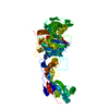 3pbqS S: Starting model for refinement |
|---|---|
| Similar structure data |
-Links
 PDBj
PDBj
- Assembly
| Deposited unit | 
| ||||||||
|---|---|---|---|---|---|---|---|---|---|
| 1 |
| ||||||||
| Unit cell |
|
-Components
| #1: Protein | Mass: 58329.453 Da / Num. of mol.: 1 Source method: isolated from a genetically manipulated source Source: (gene. exp.) Pseudomonas aeruginosa (bacteria)Gene: pbpB, ftsI, ftsI_2, ALP65_00912, CAZ10_21230, CGU42_01090, DZ934_06595, DZ962_00565, E4V10_06485, ECC04_026610, ERJ99_003095, FCG96_14995, FLI88_02250, IPC1481_11065, IPC1482_17070, IPC165_ ...Gene: pbpB, ftsI, ftsI_2, ALP65_00912, CAZ10_21230, CGU42_01090, DZ934_06595, DZ962_00565, E4V10_06485, ECC04_026610, ERJ99_003095, FCG96_14995, FLI88_02250, IPC1481_11065, IPC1482_17070, IPC165_24935, IPC170_23205, IPC669_10550, RW109_RW109_05757 Production host: References: UniProt: Q51504, UniProt: G3XD46*PLUS, serine-type D-Ala-D-Ala carboxypeptidase |
|---|---|
| #2: Chemical | ChemComp-R7G /   Mass: 688.623 Da / Num. of mol.: 1 / Source method: obtained synthetically / Formula: C27H28N8O12S / Feature type: SUBJECT OF INVESTIGATION Mass: 688.623 Da / Num. of mol.: 1 / Source method: obtained synthetically / Formula: C27H28N8O12S / Feature type: SUBJECT OF INVESTIGATION |
| #3: Water | ChemComp-HOH /  Mass: 18.015 Da / Num. of mol.: 85 / Source method: isolated from a natural source / Formula: H2O Mass: 18.015 Da / Num. of mol.: 85 / Source method: isolated from a natural source / Formula: H2O |
| Has ligand of interest | Y |
| Has protein modification | Y |
-Experimental details
-Experiment
| Experiment | Method: X-RAY DIFFRACTION / Number of used crystals: 1 |
|---|
- Sample preparation
| Crystal | Density Matthews: 2.2 Å3/Da / Density % sol: 43.98 % |
|---|---|
| Crystal grow | Temperature: 298 K / Method: vapor diffusion, sitting drop / pH: 8.5 Details: 30% PEG 4000, 0.2 M MgCl2, and 0.1 M Tris pH 8.5. Crystal was soaked for 15 minutes in 5 mM YU253434 in mother liquor. Temp details: RT |
-Data collection
| Diffraction | Mean temperature: 100 K / Serial crystal experiment: N | |||||||||||||||||||||||||||||||||||||||||||||||||||||||||||||||||||||||||||||||||||||||||||||||||||||||||||||||||||||||||||||||||||||||||||||||||||||||||||||||||||||||||||||||||||||||||||||
|---|---|---|---|---|---|---|---|---|---|---|---|---|---|---|---|---|---|---|---|---|---|---|---|---|---|---|---|---|---|---|---|---|---|---|---|---|---|---|---|---|---|---|---|---|---|---|---|---|---|---|---|---|---|---|---|---|---|---|---|---|---|---|---|---|---|---|---|---|---|---|---|---|---|---|---|---|---|---|---|---|---|---|---|---|---|---|---|---|---|---|---|---|---|---|---|---|---|---|---|---|---|---|---|---|---|---|---|---|---|---|---|---|---|---|---|---|---|---|---|---|---|---|---|---|---|---|---|---|---|---|---|---|---|---|---|---|---|---|---|---|---|---|---|---|---|---|---|---|---|---|---|---|---|---|---|---|---|---|---|---|---|---|---|---|---|---|---|---|---|---|---|---|---|---|---|---|---|---|---|---|---|---|---|---|---|---|---|---|---|---|
| Diffraction source | Source: SYNCHROTRON / Site: SSRL  / Beamline: BL9-2 / Wavelength: 0.9795 Å / Beamline: BL9-2 / Wavelength: 0.9795 Å | |||||||||||||||||||||||||||||||||||||||||||||||||||||||||||||||||||||||||||||||||||||||||||||||||||||||||||||||||||||||||||||||||||||||||||||||||||||||||||||||||||||||||||||||||||||||||||||
| Detector | Type: DECTRIS PILATUS3 6M / Detector: PIXEL / Date: Jul 5, 2019 | |||||||||||||||||||||||||||||||||||||||||||||||||||||||||||||||||||||||||||||||||||||||||||||||||||||||||||||||||||||||||||||||||||||||||||||||||||||||||||||||||||||||||||||||||||||||||||||
| Radiation | Protocol: SINGLE WAVELENGTH / Monochromatic (M) / Laue (L): M / Scattering type: x-ray | |||||||||||||||||||||||||||||||||||||||||||||||||||||||||||||||||||||||||||||||||||||||||||||||||||||||||||||||||||||||||||||||||||||||||||||||||||||||||||||||||||||||||||||||||||||||||||||
| Radiation wavelength | Wavelength: 0.9795 Å / Relative weight: 1 | |||||||||||||||||||||||||||||||||||||||||||||||||||||||||||||||||||||||||||||||||||||||||||||||||||||||||||||||||||||||||||||||||||||||||||||||||||||||||||||||||||||||||||||||||||||||||||||
| Reflection | Resolution: 2.4→50 Å / Num. obs: 20549 / % possible obs: 99.7 % / Redundancy: 8.2 % / Rmerge(I) obs: 0.136 / Rpim(I) all: 0.049 / Rrim(I) all: 0.145 / Χ2: 1.037 / Net I/σ(I): 10.9 / Num. measured all: 167804 | |||||||||||||||||||||||||||||||||||||||||||||||||||||||||||||||||||||||||||||||||||||||||||||||||||||||||||||||||||||||||||||||||||||||||||||||||||||||||||||||||||||||||||||||||||||||||||||
| Reflection shell | Diffraction-ID: 1
|
- Processing
| Software |
| ||||||||||||||||||||||||||||||||||||||||||||||||||||||||||||
|---|---|---|---|---|---|---|---|---|---|---|---|---|---|---|---|---|---|---|---|---|---|---|---|---|---|---|---|---|---|---|---|---|---|---|---|---|---|---|---|---|---|---|---|---|---|---|---|---|---|---|---|---|---|---|---|---|---|---|---|---|---|
| Refinement | Method to determine structure: MOLECULAR REPLACEMENT Starting model: 3PBQ Resolution: 2.4→39.59 Å / Cor.coef. Fo:Fc: 0.947 / Cor.coef. Fo:Fc free: 0.915 / SU B: 8.111 / SU ML: 0.188 / Cross valid method: THROUGHOUT / σ(F): 0 / ESU R: 0.464 / ESU R Free: 0.26 Details: HYDROGENS HAVE BEEN ADDED IN THE RIDING POSITIONS U VALUES : REFINED INDIVIDUALLY
| ||||||||||||||||||||||||||||||||||||||||||||||||||||||||||||
| Solvent computation | Ion probe radii: 0.8 Å / Shrinkage radii: 0.8 Å / VDW probe radii: 1.2 Å | ||||||||||||||||||||||||||||||||||||||||||||||||||||||||||||
| Displacement parameters | Biso max: 112.6 Å2 / Biso mean: 34.263 Å2 / Biso min: 13.59 Å2
| ||||||||||||||||||||||||||||||||||||||||||||||||||||||||||||
| Refinement step | Cycle: final / Resolution: 2.4→39.59 Å
| ||||||||||||||||||||||||||||||||||||||||||||||||||||||||||||
| Refine LS restraints |
| ||||||||||||||||||||||||||||||||||||||||||||||||||||||||||||
| LS refinement shell | Resolution: 2.402→2.464 Å / Rfactor Rfree error: 0 / Total num. of bins used: 20
|
