 Movie
Movie Controller

[English] 日本語
 Yorodumi
- PDB-6y6u: Structure of Pseudomonas aeruginosa Penicillin-Binding Protein 3 ... -
+ Open data

- Basic information
| Entry | Database: PDB / ID: 6y6u | ||||||
|---|---|---|---|---|---|---|---|
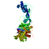
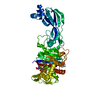
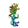
| Title | Structure of Pseudomonas aeruginosa Penicillin-Binding Protein 3 (PBP3) in complex with Compound 6 | ||||||
 Components Components | Peptidoglycan D,D-transpeptidase FtsI | ||||||
 Keywords Keywords | HYDROLASE / PBP3 / Penicillin Binding Proteins / Peptidoglycan production / transpeptidation | ||||||
| Function / homology |  Function and homology information Function and homology informationpeptidoglycan glycosyltransferase activity / serine-type D-Ala-D-Ala carboxypeptidase / serine-type D-Ala-D-Ala carboxypeptidase activity / division septum assembly / FtsZ-dependent cytokinesis / penicillin binding / peptidoglycan biosynthetic process / cell wall organization / regulation of cell shape / proteolysis / plasma membrane Similarity search - Function | ||||||
| Biological species |   Pseudomonas aeruginosa (bacteria) Pseudomonas aeruginosa (bacteria) | ||||||
| Method |  X-RAY DIFFRACTION / SYNCHROTRON / MOLECULAR REPLACEMENT / molecular replacement / Resolution: 1.55 Å X-RAY DIFFRACTION / SYNCHROTRON / MOLECULAR REPLACEMENT / molecular replacement / Resolution: 1.55 Å | ||||||
 Authors Authors | Newman, H. / Bellini, D. / Dowson, C.G. | ||||||
| Funding support |  United Kingdom, 1items United Kingdom, 1items
| ||||||
 Citation Citation | Journal: Chem Sci / Year: 2020 Title: Demonstration of the utility of DOS-derived fragment libraries for rapid hit derivatisation in a multidirectional fashion Authors: Kidd, S.L. / Fowler, E. / Reinhardt, T. / Compton, T. / Mateu, N. / Newman, H. / Bellini, D. / Talon, R. / McLoughlin, J. / Krojer, T. / Aimon, A. / Bradley, A. / Fairhead, M. / Brear, P. / ...Authors: Kidd, S.L. / Fowler, E. / Reinhardt, T. / Compton, T. / Mateu, N. / Newman, H. / Bellini, D. / Talon, R. / McLoughlin, J. / Krojer, T. / Aimon, A. / Bradley, A. / Fairhead, M. / Brear, P. / Diaz-Saez, L. / McAuley, K. / Sore, H.F. / Madin, A. / O'Donovan, D.H. / Huber, K.V.M. / Hyvonen, M. / Dowson, C.G. / Spring, D.R. | ||||||
| History |
|
- Structure visualization
| Structure viewer | Molecule: MolmilJmol/JSmol |
|---|
- Downloads & links
-Download
| PDBx/mmCIF format | 6y6u.cif.gz | 228.8 KB | Display | PDBx/mmCIF format |
|---|---|---|---|---|
| PDB format | pdb6y6u.ent.gz | 180.5 KB | Display | PDB format |
| PDBx/mmJSON format | 6y6u.json.gz | Tree view | PDBx/mmJSON format | |
| Others |  Other downloads Other downloads |
-Validation report
| Arichive directory | https://data.pdbj.org/pub/pdb/validation_reports/y6/6y6uftp://data.pdbj.org/pub/pdb/validation_reports/y6/6y6u | HTTPS FTP |
|---|
-Related structure data
| Related structure data |  6y6nC  6y6oC  6hzrS S: Starting model for refinement C: citing same article ( |
|---|---|
| Similar structure data |


-Links
 PDBj
PDBj
- Assembly
| Deposited unit | 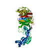
| ||||||||
|---|---|---|---|---|---|---|---|---|---|
| 1 |
| ||||||||
| Unit cell |
|
-Components
| #1: Protein | Mass: 56431.324 Da / Num. of mol.: 1 Source method: isolated from a genetically manipulated source Source: (gene. exp.) Pseudomonas aeruginosa (strain ATCC 15692 / DSM 22644 / CIP 104116 / JCM 14847 / LMG 12228 / 1C / PRS 101 / PAO1) (bacteria)Gene: ftsI, pbpB, PA4418 / Production host: References: UniProt: G3XD46, serine-type D-Ala-D-Ala carboxypeptidase | ||||||||
|---|---|---|---|---|---|---|---|---|---|
| #2: Chemical | ChemComp-GOL /   Mass: 92.094 Da / Num. of mol.: 5 / Source method: obtained synthetically / Formula: C3H8O3 Mass: 92.094 Da / Num. of mol.: 5 / Source method: obtained synthetically / Formula: C3H8O3#3: Chemical | ChemComp-ODZ / |   Mass: 263.289 Da / Num. of mol.: 1 / Source method: obtained synthetically / Formula: C14H17NO4 / Feature type: SUBJECT OF INVESTIGATION Mass: 263.289 Da / Num. of mol.: 1 / Source method: obtained synthetically / Formula: C14H17NO4 / Feature type: SUBJECT OF INVESTIGATION#4: Water | ChemComp-HOH / |  Mass: 18.015 Da / Num. of mol.: 348 / Source method: isolated from a natural source / Formula: H2O Mass: 18.015 Da / Num. of mol.: 348 / Source method: isolated from a natural source / Formula: H2OHas ligand of interest | Y | Has protein modification | Y | |
-Experimental details
-Experiment
| Experiment | Method: X-RAY DIFFRACTION / Number of used crystals: 1 |
|---|
- Sample preparation
| Crystal | Density Matthews: 2.27 Å3/Da / Density % sol: 45.92 % / Mosaicity: 0.08 ° |
|---|---|
| Crystal grow | Temperature: 293 K / Method: vapor diffusion, sitting drop / pH: 7.8 Details: 25% (w/v) polyethylene glycol 3 350, 0.1 M Bis-Tris propane pH 7.8 and 1% (w/v) protamine sulphate |
-Data collection
| Diffraction | Mean temperature: 100 K / Serial crystal experiment: N |
|---|---|
| Diffraction source | Source: SYNCHROTRON / Site: Diamond / Beamline: I03 / Wavelength: 0.9793 Å |
| Detector | Type: DECTRIS PILATUS3 6M / Detector: PIXEL / Date: Jul 13, 2018 / Details: mirrors |
| Radiation | Monochromator: silicon / Protocol: SINGLE WAVELENGTH / Monochromatic (M) / Laue (L): M / Scattering type: x-ray |
| Radiation wavelength | Wavelength: 0.9793 Å / Relative weight: 1 |
| Reflection | Resolution: 1.55→61.09 Å / Num. obs: 56715 / % possible obs: 95.5 % / Redundancy: 7.9 % / CC1/2: 0.999 / Rmerge(I) obs: 0.051 / Rpim(I) all: 0.02 / Net I/σ(I): 17.5 |
| Reflection shell | Resolution: 1.55→1.69 Å / Redundancy: 7.7 % / Rmerge(I) obs: 1.179 / Mean I/σ(I) obs: 1.6 / Num. unique obs: 2836 / CC1/2: 0.705 / Rpim(I) all: 0.448 / % possible all: 64.7 |
-Phasing
| Phasing | Method: molecular replacement |
|---|
- Processing
| Software |
| |||||||||||||||||||||||||||||||||||||||||||||||||||||||||||||||||
|---|---|---|---|---|---|---|---|---|---|---|---|---|---|---|---|---|---|---|---|---|---|---|---|---|---|---|---|---|---|---|---|---|---|---|---|---|---|---|---|---|---|---|---|---|---|---|---|---|---|---|---|---|---|---|---|---|---|---|---|---|---|---|---|---|---|---|
| Refinement | Method to determine structure: MOLECULAR REPLACEMENT Starting model: 6HZR Resolution: 1.55→61.09 Å / Cor.coef. Fo:Fc: 0.975 / Cor.coef. Fo:Fc free: 0.95 / SU B: 10.417 / SU ML: 0.137 / SU R Cruickshank DPI: 0.1403 / Cross valid method: FREE R-VALUE / σ(F): 0 / ESU R: 0.14 / ESU R Free: 0.111 Details: HYDROGENS HAVE BEEN ADDED IN THE RIDING POSITIONS U VALUES : REFINED INDIVIDUALLY
| |||||||||||||||||||||||||||||||||||||||||||||||||||||||||||||||||
| Solvent computation | Ion probe radii: 0.8 Å / Shrinkage radii: 0.8 Å / VDW probe radii: 1.2 Å | |||||||||||||||||||||||||||||||||||||||||||||||||||||||||||||||||
| Displacement parameters | Biso max: 450.53 Å2 / Biso mean: 38.566 Å2 / Biso min: 19.03 Å2
| |||||||||||||||||||||||||||||||||||||||||||||||||||||||||||||||||
| Refinement step | Cycle: final / Resolution: 1.55→61.09 Å
| |||||||||||||||||||||||||||||||||||||||||||||||||||||||||||||||||
| Refine LS restraints |
| |||||||||||||||||||||||||||||||||||||||||||||||||||||||||||||||||
| LS refinement shell | Resolution: 1.552→1.592 Å / Rfactor Rfree error: 0 / Total num. of bins used: 20
|
