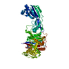
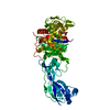
Entry Database : PDB / ID : 6hr9Title Nitrocefin acylation of both catalytic serines of the Y409 mutant of penicillin-binding protein 3 from P. aeruginosa Peptidoglycan D,D-transpeptidase FtsI Keywords / / Function / homology Function Domain/homology Component
/ / / / / / / / / / / / / / / / / / / / / / / / / / / / / / Biological species Pseudomonas aeruginosa (bacteria)Method / / / Resolution : 1.99 Å Authors Bellini, D. / Dowson, C.G. Funding support Organization Grant number Country Medical Research Council (MRC, United Kingdom) grant.MR/P007503/1
Journal : To Be Published Title : Nitrocefin acylation of both catalytic serines of penicillin-binding protein 3 from P. aeruginosaAuthors : Bellini, D. / Dowson, C.G. History Deposition Sep 26, 2018 Deposition site / Processing site Revision 1.0 Oct 23, 2019 Provider / Type Revision 2.0 Aug 25, 2021 Group Advisory / Atomic model ... Advisory / Atomic model / Author supporting evidence / Data collection / Database references / Derived calculations / Other / Refinement description / Structure summary Category atom_site / atom_sites ... atom_site / atom_sites / database_2 / database_PDB_caveat / diffrn / pdbx_audit_support / pdbx_entity_instance_feature / pdbx_entry_details / pdbx_nonpoly_scheme / pdbx_poly_seq_scheme / pdbx_struct_sheet_hbond / pdbx_unobs_or_zero_occ_residues / pdbx_validate_chiral / pdbx_validate_close_contact / pdbx_validate_planes / pdbx_validate_rmsd_angle / pdbx_validate_rmsd_bond / pdbx_validate_symm_contact / pdbx_validate_torsion / refine / refine_hist / refine_ls_restr / refine_ls_shell / reflns / reflns_shell / software / struct_conf / struct_conn / struct_mon_prot_cis / struct_sheet / struct_sheet_order / struct_sheet_range / struct_site / struct_site_gen Item _atom_sites.fract_transf_matrix[2][1] / _atom_sites.fract_transf_matrix[3][2] ... _atom_sites.fract_transf_matrix[2][1] / _atom_sites.fract_transf_matrix[3][2] / _database_2.pdbx_DOI / _database_2.pdbx_database_accession / _diffrn.pdbx_serial_crystal_experiment / _pdbx_audit_support.funding_organization / _pdbx_nonpoly_scheme.auth_mon_id / _pdbx_nonpoly_scheme.auth_seq_num / _pdbx_poly_seq_scheme.auth_mon_id / _pdbx_poly_seq_scheme.auth_seq_num / _pdbx_poly_seq_scheme.pdb_mon_id / _refine.B_iso_max / _refine.B_iso_mean / _refine.B_iso_min / _refine.aniso_B[1][1] / _refine.aniso_B[1][2] / _refine.aniso_B[2][3] / _refine.aniso_B[3][3] / _refine.correlation_coeff_Fo_to_Fc / _refine.correlation_coeff_Fo_to_Fc_free / _refine.details / _refine.ls_R_factor_R_free / _refine.ls_R_factor_R_work / _refine.ls_R_factor_obs / _refine.overall_SU_B / _refine.overall_SU_ML / _refine.pdbx_ls_sigma_F / _refine.pdbx_method_to_determine_struct / _refine.pdbx_overall_ESU_R / _refine.pdbx_overall_ESU_R_Free / _refine.pdbx_starting_model / _refine.pdbx_stereochemistry_target_values / _refine.solvent_model_details / _refine_hist.cycle_id / _refine_hist.number_atoms_total / _refine_hist.pdbx_B_iso_mean_ligand / _refine_hist.pdbx_B_iso_mean_solvent / _refine_hist.pdbx_number_atoms_ligand / _refine_hist.pdbx_number_atoms_protein / _refine_hist.pdbx_number_residues_total / _refine_ls_shell.R_factor_R_free / _refine_ls_shell.R_factor_R_free_error / _refine_ls_shell.R_factor_R_work / _refine_ls_shell.number_reflns_all / _reflns.B_iso_Wilson_estimate / _reflns_shell.number_unique_obs / _reflns_shell.pdbx_CC_half / _software.version / _struct_conf.beg_auth_comp_id / _struct_conf.beg_auth_seq_id / _struct_conf.beg_label_comp_id / _struct_conf.beg_label_seq_id / _struct_conf.end_auth_comp_id / _struct_conf.end_auth_seq_id / _struct_conf.end_label_comp_id / _struct_conf.end_label_seq_id / _struct_conf.pdbx_PDB_helix_length / _struct_conn.pdbx_dist_value / _struct_mon_prot_cis.pdbx_omega_angle / _struct_sheet.number_strands Description / Details / Provider / Type Revision 3.0 Jul 20, 2022 Group Advisory / Atomic model ... Advisory / Atomic model / Data collection / Derived calculations / Non-polymer description / Refinement description / Structure summary Category atom_site / chem_comp ... atom_site / chem_comp / database_PDB_caveat / diffrn_radiation_wavelength / diffrn_source / entity / pdbx_nonpoly_scheme / pdbx_poly_seq_scheme / pdbx_struct_assembly_prop / pdbx_struct_sheet_hbond / pdbx_unobs_or_zero_occ_residues / pdbx_validate_chiral / pdbx_validate_close_contact / pdbx_validate_peptide_omega / pdbx_validate_rmsd_angle / pdbx_validate_rmsd_bond / pdbx_validate_symm_contact / pdbx_validate_torsion / refine / refine_hist / refine_ls_restr / refine_ls_shell / software / struct_conf / struct_conn / struct_mon_prot_cis / struct_sheet_range Item _chem_comp.formula / _chem_comp.formula_weight ... _chem_comp.formula / _chem_comp.formula_weight / _chem_comp.pdbx_synonyms / _diffrn_radiation_wavelength.wavelength / _diffrn_source.pdbx_wavelength_list / _entity.formula_weight / _entity.pdbx_mutation / _entity.pdbx_number_of_molecules / _pdbx_poly_seq_scheme.auth_mon_id / _pdbx_poly_seq_scheme.auth_seq_num / _pdbx_poly_seq_scheme.pdb_mon_id / _pdbx_struct_assembly_prop.value / _pdbx_struct_sheet_hbond.range_1_auth_comp_id / _pdbx_struct_sheet_hbond.range_1_auth_seq_id / _pdbx_struct_sheet_hbond.range_1_label_comp_id / _pdbx_struct_sheet_hbond.range_1_label_seq_id / _pdbx_struct_sheet_hbond.range_2_auth_comp_id / _pdbx_struct_sheet_hbond.range_2_auth_seq_id / _pdbx_struct_sheet_hbond.range_2_label_comp_id / _pdbx_struct_sheet_hbond.range_2_label_seq_id / _refine.B_iso_max / _refine.B_iso_mean / _refine.B_iso_min / _refine.aniso_B[1][1] / _refine.aniso_B[2][2] / _refine.aniso_B[3][3] / _refine.correlation_coeff_Fo_to_Fc / _refine.correlation_coeff_Fo_to_Fc_free / _refine.ls_R_factor_R_free / _refine.ls_R_factor_R_work / _refine.ls_R_factor_obs / _refine.overall_SU_B / _refine.overall_SU_ML / _refine.pdbx_overall_ESU_R / _refine.pdbx_overall_ESU_R_Free / _refine_hist.number_atoms_solvent / _refine_hist.number_atoms_total / _refine_hist.pdbx_B_iso_mean_ligand / _refine_hist.pdbx_B_iso_mean_solvent / _refine_hist.pdbx_number_atoms_protein / _refine_hist.pdbx_number_residues_total / _refine_ls_restr.dev_ideal / _refine_ls_restr.dev_ideal_target / _refine_ls_restr.number / _refine_ls_shell.R_factor_R_free / _refine_ls_shell.R_factor_R_work / _software.version / _struct_conf.beg_auth_comp_id / _struct_conf.beg_auth_seq_id / _struct_conf.beg_label_comp_id / _struct_conf.beg_label_seq_id / _struct_conf.end_auth_comp_id / _struct_conf.end_auth_seq_id / _struct_conf.end_label_comp_id / _struct_conf.end_label_seq_id / _struct_conf.pdbx_PDB_helix_class / _struct_conf.pdbx_PDB_helix_length / _struct_conn.pdbx_dist_value / _struct_conn.pdbx_leaving_atom_flag / _struct_conn.ptnr2_auth_seq_id / _struct_mon_prot_cis.pdbx_omega_angle / _struct_sheet_range.beg_auth_comp_id / _struct_sheet_range.beg_auth_seq_id / _struct_sheet_range.beg_label_comp_id / _struct_sheet_range.beg_label_seq_id / _struct_sheet_range.end_auth_comp_id / _struct_sheet_range.end_auth_seq_id / _struct_sheet_range.end_label_comp_id / _struct_sheet_range.end_label_seq_id Description / Provider / Type Revision 3.1 Jan 24, 2024 Group / Refinement descriptionCategory / chem_comp_bond / pdbx_initial_refinement_modelRevision 3.2 Nov 20, 2024 Group / Category / pdbx_modification_feature / Item
Show all Show less
 Movie
Movie Controller

 Yorodumi
 Open data
 Basic information
 Components
 Keywords
 Function and homology information
 Pseudomonas aeruginosa (bacteria)
 X-RAY DIFFRACTION /
  Authors
 United Kingdom, 1items
  Citation
 Structure visualization
 Downloads & links
 Other downloads


 PDBj
PDBj
 Assembly

 Mass: 534.519 Da / Num. of mol.: 2 / Source method: obtained synthetically / Formula: C21H18N4O9S2 / Feature type: SUBJECT OF INVESTIGATION
 Mass: 18.015 Da / Num. of mol.: 102 / Source method: isolated from a natural source / Formula: H2O
 Sample preparation
 Processing