 Movie
Movie Controller

[English] 日本語
 Yorodumi
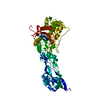
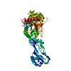
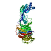
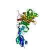
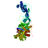
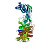
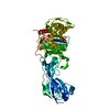
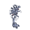
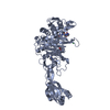
- PDB-6hr6: Nitrocefin reacted with catalytic serine (Ser294) of penicillin-b... -
+ Open data

- Basic information
| Entry | Database: PDB / ID: 6hr6 | ||||||||||||
|---|---|---|---|---|---|---|---|---|---|---|---|---|---|
| Title | Nitrocefin reacted with catalytic serine (Ser294) of penicillin-binding protein 3 from Pseudomonas aeruginosa | ||||||||||||
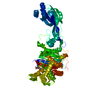
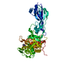
 Components Components | Peptidoglycan D,D-transpeptidase FtsI | ||||||||||||
 Keywords Keywords | PENICILLIN-BINDING PROTEIN / penicillin-binding protein peptidoglycan | ||||||||||||
| Function / homology |  Function and homology information Function and homology informationpeptidoglycan glycosyltransferase activity / serine-type D-Ala-D-Ala carboxypeptidase / serine-type D-Ala-D-Ala carboxypeptidase activity / division septum assembly / FtsZ-dependent cytokinesis / penicillin binding / peptidoglycan biosynthetic process / cell wall organization / regulation of cell shape / proteolysis / plasma membrane Similarity search - Function | ||||||||||||
| Biological species |   Pseudomonas aeruginosa (bacteria) Pseudomonas aeruginosa (bacteria) | ||||||||||||
| Method |  X-RAY DIFFRACTION / SYNCHROTRON / MOLECULAR REPLACEMENT / Resolution: 2.53 Å X-RAY DIFFRACTION / SYNCHROTRON / MOLECULAR REPLACEMENT / Resolution: 2.53 Å | ||||||||||||
 Authors Authors | Bellini, D. / Dowson, C.G. | ||||||||||||
| Funding support |  United Kingdom, 1items United Kingdom, 1items
| ||||||||||||
 Citation Citation | Journal: To Be Published Title: Nitrocefin acylation of single catalytic serine of penicillin-binding protein 3 from P. aeruginosa Authors: Bellini, D. / Dowson, C.G. | ||||||||||||
| History |
|
- Structure visualization
| Structure viewer | Molecule: MolmilJmol/JSmol |
|---|
- Downloads & links
-Download
| PDBx/mmCIF format | 6hr6.cif.gz | 114.4 KB | Display | PDBx/mmCIF format |
|---|---|---|---|---|
| PDB format | pdb6hr6.ent.gz | 83 KB | Display | PDB format |
| PDBx/mmJSON format | 6hr6.json.gz | Tree view | PDBx/mmJSON format | |
| Others |  Other downloads Other downloads |
-Validation report
| Arichive directory | https://data.pdbj.org/pub/pdb/validation_reports/hr/6hr6ftp://data.pdbj.org/pub/pdb/validation_reports/hr/6hr6 | HTTPS FTP |
|---|
-Related structure data
| Related structure data | 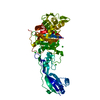 3oc2S S: Starting model for refinement |
|---|---|
| Similar structure data |

-Links
 PDBj
PDBj
- Assembly
| Deposited unit | 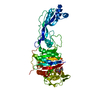
| ||||||||
|---|---|---|---|---|---|---|---|---|---|
| 1 |
| ||||||||
| Unit cell |
|
-Components
| #1: Protein | Mass: 57872.883 Da / Num. of mol.: 1 Source method: isolated from a genetically manipulated source Source: (gene. exp.) Pseudomonas aeruginosa (bacteria)Strain: ATCC 15692 / DSM 22644 / CIP 104116 / JCM 14847 / LMG 12228 / 1C / PRS 101 / PAO1 Gene: ftsI, pbpB, PA4418 / Production host: References: UniProt: G3XD46, serine-type D-Ala-D-Ala carboxypeptidase |
|---|---|
| #2: Chemical | ChemComp-NEF /   Mass: 534.519 Da / Num. of mol.: 1 / Source method: obtained synthetically / Formula: C21H18N4O9S2 / Feature type: SUBJECT OF INVESTIGATION Mass: 534.519 Da / Num. of mol.: 1 / Source method: obtained synthetically / Formula: C21H18N4O9S2 / Feature type: SUBJECT OF INVESTIGATION |
| #3: Water | ChemComp-HOH /  Mass: 18.015 Da / Num. of mol.: 20 / Source method: isolated from a natural source / Formula: H2O Mass: 18.015 Da / Num. of mol.: 20 / Source method: isolated from a natural source / Formula: H2O |
| Has ligand of interest | Y |
| Has protein modification | Y |
-Experimental details
-Experiment
| Experiment | Method: X-RAY DIFFRACTION / Number of used crystals: 1 |
|---|
- Sample preparation
| Crystal | Density Matthews: 2.18 Å3/Da / Density % sol: 43.64 % |
|---|---|
| Crystal grow | Temperature: 294 K / Method: vapor diffusion, sitting drop Details: 25%(w/v) polyethylene glycol 3350, 0.1M Bis-Tris propane, 1%(w/v) protamine sulphate, pH 7.8. |
-Data collection
| Diffraction | Mean temperature: 100 K / Serial crystal experiment: N |
|---|---|
| Diffraction source | Source: SYNCHROTRON / Site: Diamond / Beamline: I04 / Wavelength: 0.9795 Å |
| Detector | Type: DECTRIS PILATUS 6M-F / Detector: PIXEL / Date: Feb 4, 2018 |
| Radiation | Protocol: SINGLE WAVELENGTH / Monochromatic (M) / Laue (L): M / Scattering type: x-ray |
| Radiation wavelength | Wavelength: 0.9795 Å / Relative weight: 1 |
| Reflection | Resolution: 2.53→53 Å / Num. obs: 17652 / % possible obs: 100 % / Redundancy: 8.5 % / Biso Wilson estimate: 46.48 Å2 / Rrim(I) all: 0.191 / Net I/σ(I): 7.8 |
| Reflection shell | Resolution: 2.53→2.57 Å / Num. unique obs: 1256 / Rpim(I) all: 0.963 |
- Processing
| Software |
| |||||||||||||||||||||||||||||||||||||||||||||||||
|---|---|---|---|---|---|---|---|---|---|---|---|---|---|---|---|---|---|---|---|---|---|---|---|---|---|---|---|---|---|---|---|---|---|---|---|---|---|---|---|---|---|---|---|---|---|---|---|---|---|---|
| Refinement | Method to determine structure: MOLECULAR REPLACEMENT Starting model: 3OC2 Resolution: 2.53→45.54 Å / SU ML: 0.46 / Cross valid method: THROUGHOUT / σ(F): 1.33 / Phase error: 33.28 / Stereochemistry target values: ML
| |||||||||||||||||||||||||||||||||||||||||||||||||
| Solvent computation | Shrinkage radii: 0.9 Å / VDW probe radii: 1.11 Å / Solvent model: FLAT BULK SOLVENT MODEL | |||||||||||||||||||||||||||||||||||||||||||||||||
| Displacement parameters | Biso max: 145.05 Å2 / Biso mean: 56.4722 Å2 / Biso min: 20.09 Å2 | |||||||||||||||||||||||||||||||||||||||||||||||||
| Refinement step | Cycle: final / Resolution: 2.53→45.54 Å
| |||||||||||||||||||||||||||||||||||||||||||||||||
| LS refinement shell | Refine-ID: X-RAY DIFFRACTION / Rfactor Rfree error: 0 / Total num. of bins used: 6
|
