- PDB-6y6o: Structure of mature activin A with small molecule 42 -
+
Open data
ID or keywords:
Loading...
-
Basic information
Entry
Database: PDB / ID: 6y6o
Title
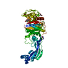
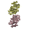
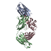
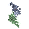
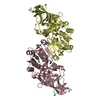
Structure of mature activin A with small molecule 42
Components
Inhibin beta A chain
Keywords
SIGNALING PROTEIN / activin A / TGF-beta / growth factor / FBDD / inhibitor
Function / homology
Function and homology information
activin A complex / inhibin A complex / cardiac fibroblast cell development / androst-4-ene-3,17-dione biosynthetic process / negative regulation of B cell differentiation / regulation of follicle-stimulating hormone secretion / GABAergic neuron differentiation / Antagonism of Activin by Follistatin / TGFBR3 regulates activin signaling / negative regulation of follicle-stimulating hormone secretion ...activin A complex / inhibin A complex / cardiac fibroblast cell development / androst-4-ene-3,17-dione biosynthetic process / negative regulation of B cell differentiation / regulation of follicle-stimulating hormone secretion / GABAergic neuron differentiation / Antagonism of Activin by Follistatin / TGFBR3 regulates activin signaling / negative regulation of follicle-stimulating hormone secretion / positive regulation of ovulation / type II activin receptor binding / progesterone secretion / Sertoli cell differentiation / striatal medium spiny neuron differentiation / Glycoprotein hormones / negative regulation of macrophage differentiation / negative regulation of phosphorylation / positive regulation of follicle-stimulating hormone secretion / hemoglobin biosynthetic process / cellular response to oxygen-glucose deprivation / testosterone biosynthetic process / cellular response to follicle-stimulating hormone stimulus / cellular response to cholesterol / Signaling by BMP / SMAD protein signal transduction / Signaling by Activin / activin receptor signaling pathway / positive regulation of extrinsic apoptotic signaling pathway in absence of ligand / response to aldosterone / mesodermal cell differentiation / odontogenesis / cell surface receptor protein serine/threonine kinase signaling pathway / positive regulation of transcription by RNA polymerase III / negative regulation of G1/S transition of mitotic cell cycle / eyelid development in camera-type eye / endodermal cell differentiation / negative regulation of type II interferon production / androgen metabolic process / roof of mouth development / positive regulation of collagen biosynthetic process / positive regulation of protein metabolic process / peptide hormone binding / cellular response to angiotensin / positive regulation of SMAD protein signal transduction / hair follicle development / hematopoietic progenitor cell differentiation / ovarian follicle development / extrinsic apoptotic signaling pathway / positive regulation of erythrocyte differentiation / cytokine activity / erythrocyte differentiation / growth factor activity / defense response / hormone activity / negative regulation of cell growth / male gonad development / autophagy / cytokine-mediated signaling pathway / cell-cell signaling / nervous system development / transcription by RNA polymerase II / cellular response to hypoxia / cell differentiation / positive regulation of ERK1 and ERK2 cascade / cell surface receptor signaling pathway / negative regulation of cell population proliferation / positive regulation of gene expression / regulation of transcription by RNA polymerase II / positive regulation of DNA-templated transcription / protein-containing complex binding / perinuclear region of cytoplasm / positive regulation of transcription by RNA polymerase II / : / extracellular region / identical protein binding Similarity search - Function
Inhibin, beta A subunit / TGF-beta, propeptide / TGF-beta propeptide / Transforming growth factor beta, conserved site / TGF-beta family signature. / Transforming growth factor-beta-related / Transforming growth factor-beta (TGF-beta) family / Transforming growth factor-beta, C-terminal / Transforming growth factor beta like domain / TGF-beta family profile. / Cystine-knot cytokine Similarity search - Domain/homology
In the structure databanks used in Yorodumi, some data are registered as the other names, "COVID-19 virus" and "2019-nCoV". Here are the details of the virus and the list of structure data.
Jan 31, 2019. EMDB accession codes are about to change! (news from PDBe EMDB page)
EMDB accession codes are about to change! (news from PDBe EMDB page)
The allocation of 4 digits for EMDB accession codes will soon come to an end. Whilst these codes will remain in use, new EMDB accession codes will include an additional digit and will expand incrementally as the available range of codes is exhausted. The current 4-digit format prefixed with “EMD-” (i.e. EMD-XXXX) will advance to a 5-digit format (i.e. EMD-XXXXX), and so on. It is currently estimated that the 4-digit codes will be depleted around Spring 2019, at which point the 5-digit format will come into force.
The EM Navigator/Yorodumi systems omit the EMD- prefix.
Related info.:Q: What is EMD? / ID/Accession-code notation in Yorodumi/EM Navigator
Yorodumi is a browser for structure data from EMDB, PDB, SASBDB, etc.
This page is also the successor to EM Navigator detail page, and also detail information page/front-end page for Omokage search.
The word "yorodu" (or yorozu) is an old Japanese word meaning "ten thousand". "mi" (miru) is to see.
Related info.:EMDB / PDB / SASBDB / Comparison of 3 databanks / Yorodumi Search / Aug 31, 2016. New EM Navigator & Yorodumi / Yorodumi Papers / Jmol/JSmol / Function and homology information / Changes in new EM Navigator and Yorodumi
 Movie
Movie Controller


 Open data
 Basic information
 Components
 Keywords
 Function and homology information
 Homo sapiens (human)
 X-RAY DIFFRACTION /
  Authors
 United Kingdom, 1items
  Citation
 Structure visualization
 Downloads & links
 Other downloads


 PDBj
PDBj


 Assembly
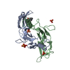


 Mass: 96.063 Da / Num. of mol.: 5 / Source method: obtained synthetically / Formula: SO4
 Mass: 260.331 Da / Num. of mol.: 1 / Source method: obtained synthetically / Formula: C15H20N2O2 / Feature type: SUBJECT OF INVESTIGATION
 Mass: 18.015 Da / Num. of mol.: 49 / Source method: isolated from a natural source / Formula: H2O
 Sample preparation
 Processing