 Movie
Movie Controller

[English] 日本語
 Yorodumi
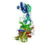
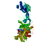
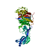
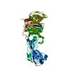
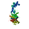
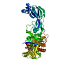
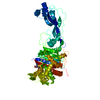
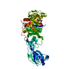
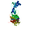
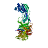
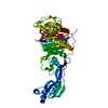
- PDB-4oom: Crystal structure of PBP3 in complex with BAL30072 ((2Z)-2-(2-ami... -
+ Open data

- Basic information
| Entry | Database: PDB / ID: 4oom | ||||||
|---|---|---|---|---|---|---|---|
| Title | Crystal structure of PBP3 in complex with BAL30072 ((2Z)-2-(2-amino-1,3-thiazol-4-yl)-2-{[(1,5-dihydroxy-4-oxo-1,4-dihydropyridin-2-yl)methoxy]imino}-N-{(2S)-1-hydroxy-3-methyl-3-[(sulfooxy)amino]butan-2-yl}ethanamide) | ||||||
 Components Components | Cell division protein FtsI [Peptidoglycan synthetase] | ||||||
 Keywords Keywords | transferase/transferase inhibitor / PBP3 / transferase-transferase inhibitor complex | ||||||
| Function / homology | Beta-Lactamase - #330 / Beta-Lactamase / Beta-lactamase / DD-peptidase/beta-lactamase superfamily / 2-Layer Sandwich / 3-Layer(aba) Sandwich / Alpha Beta / Chem-2U3 / :  Function and homology information Function and homology information | ||||||
| Biological species |  Pseudomonas aeruginosa PA1R (bacteria) Pseudomonas aeruginosa PA1R (bacteria) | ||||||
| Method |  X-RAY DIFFRACTION / SYNCHROTRON / MOLECULAR REPLACEMENT / Resolution: 2 Å X-RAY DIFFRACTION / SYNCHROTRON / MOLECULAR REPLACEMENT / Resolution: 2 Å | ||||||
 Authors Authors | Han, S. / Caspers, N. / Knafels, J.D. | ||||||
 Citation Citation | Journal: J.Med.Chem. / Year: 2014 Title: Siderophore receptor-mediated uptake of lactivicin analogues in gram-negative bacteria. Authors: Starr, J. / Brown, M.F. / Aschenbrenner, L. / Caspers, N. / Che, Y. / Gerstenberger, B.S. / Huband, M. / Knafels, J.D. / Lemmon, M.M. / Li, C. / McCurdy, S.P. / McElroy, E. / Rauckhorst, M.R. ...Authors: Starr, J. / Brown, M.F. / Aschenbrenner, L. / Caspers, N. / Che, Y. / Gerstenberger, B.S. / Huband, M. / Knafels, J.D. / Lemmon, M.M. / Li, C. / McCurdy, S.P. / McElroy, E. / Rauckhorst, M.R. / Tomaras, A.P. / Young, J.A. / Zaniewski, R.P. / Shanmugasundaram, V. / Han, S. | ||||||
| History |
|
- Structure visualization
| Structure viewer | Molecule: MolmilJmol/JSmol |
|---|
- Downloads & links
-Download
| PDBx/mmCIF format | 4oom.cif.gz | 208.7 KB | Display | PDBx/mmCIF format |
|---|---|---|---|---|
| PDB format | pdb4oom.ent.gz | 164.6 KB | Display | PDB format |
| PDBx/mmJSON format | 4oom.json.gz | Tree view | PDBx/mmJSON format | |
| Others |  Other downloads Other downloads |
-Validation report
| Arichive directory | https://data.pdbj.org/pub/pdb/validation_reports/oo/4oomftp://data.pdbj.org/pub/pdb/validation_reports/oo/4oom | HTTPS FTP |
|---|
-Related structure data


-Links
 PDBj
PDBj- Assembly
| Deposited unit | 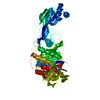
| ||||||||
|---|---|---|---|---|---|---|---|---|---|
| 1 |
| ||||||||
| Unit cell |
|
-Components
| #1: Protein | Mass: 58329.453 Da / Num. of mol.: 1 / Fragment: PBP3 Source method: isolated from a genetically manipulated source Source: (gene. exp.) Pseudomonas aeruginosa PA1R (bacteria) / Gene: PA1R_gp2318 / Production host: References: UniProt: U6AVY0, peptidoglycan glycosyltransferase |
|---|---|
| #2: Chemical | ChemComp-2U3 / (  Mass: 522.510 Da / Num. of mol.: 1 / Source method: obtained synthetically / Formula: C16H22N6O10S2 Mass: 522.510 Da / Num. of mol.: 1 / Source method: obtained synthetically / Formula: C16H22N6O10S2 |
| #3: Water | ChemComp-HOH /  Mass: 18.015 Da / Num. of mol.: 259 / Source method: isolated from a natural source / Formula: H2O Mass: 18.015 Da / Num. of mol.: 259 / Source method: isolated from a natural source / Formula: H2O |
| Has protein modification | Y |
-Experimental details
-Experiment
| Experiment | Method: X-RAY DIFFRACTION / Number of used crystals: 1 |
|---|
- Sample preparation
| Crystal | Density Matthews: 2.12 Å3/Da / Density % sol: 42.01 % |
|---|---|
| Crystal grow | Temperature: 294 K / Method: vapor diffusion, hanging drop / pH: 8.5 Details: 30% PEG 4K, 0.2M MgCl2, 0.1M Tris, pH 8.5, VAPOR DIFFUSION, HANGING DROP, temperature 294K |
-Data collection
| Diffraction | Mean temperature: 173 K |
|---|---|
| Diffraction source | Source: SYNCHROTRON / Site: APS  / Beamline: 17-ID / Wavelength: 1 Å / Beamline: 17-ID / Wavelength: 1 Å |
| Detector | Type: DECTRIS PILATUS 6M / Detector: PIXEL / Date: Aug 18, 2012 |
| Radiation | Monochromator: mirror / Protocol: SINGLE WAVELENGTH / Monochromatic (M) / Laue (L): M / Scattering type: x-ray |
| Radiation wavelength | Wavelength: 1 Å / Relative weight: 1 |
| Reflection | Resolution: 2→30 Å / Num. all: 148320 / Num. obs: 32905 / % possible obs: 95.8 % / Observed criterion σ(F): 1 / Observed criterion σ(I): 1 / Biso Wilson estimate: 29.82 Å2 |
| Reflection shell | Resolution: 2→2.07 Å / % possible all: 81.6 |
- Processing
| Software |
| ||||||||||||||||||||||||||||||||||||||||||||||||||||||||||||||||||||||||||||||||||||||||||||||||||||||||||||||||||
|---|---|---|---|---|---|---|---|---|---|---|---|---|---|---|---|---|---|---|---|---|---|---|---|---|---|---|---|---|---|---|---|---|---|---|---|---|---|---|---|---|---|---|---|---|---|---|---|---|---|---|---|---|---|---|---|---|---|---|---|---|---|---|---|---|---|---|---|---|---|---|---|---|---|---|---|---|---|---|---|---|---|---|---|---|---|---|---|---|---|---|---|---|---|---|---|---|---|---|---|---|---|---|---|---|---|---|---|---|---|---|---|---|---|---|---|
| Refinement | Method to determine structure: MOLECULAR REPLACEMENT / Resolution: 2→29.52 Å / Cor.coef. Fo:Fc: 0.952 / Cor.coef. Fo:Fc free: 0.9393 / SU R Cruickshank DPI: 0.188 / Cross valid method: THROUGHOUT / σ(F): 0 / Stereochemistry target values: Engh & Huber
| ||||||||||||||||||||||||||||||||||||||||||||||||||||||||||||||||||||||||||||||||||||||||||||||||||||||||||||||||||
| Displacement parameters | Biso mean: 41.74 Å2
| ||||||||||||||||||||||||||||||||||||||||||||||||||||||||||||||||||||||||||||||||||||||||||||||||||||||||||||||||||
| Refine analyze | Luzzati coordinate error obs: 0.262 Å | ||||||||||||||||||||||||||||||||||||||||||||||||||||||||||||||||||||||||||||||||||||||||||||||||||||||||||||||||||
| Refinement step | Cycle: LAST / Resolution: 2→29.52 Å
| ||||||||||||||||||||||||||||||||||||||||||||||||||||||||||||||||||||||||||||||||||||||||||||||||||||||||||||||||||
| Refine LS restraints |
| ||||||||||||||||||||||||||||||||||||||||||||||||||||||||||||||||||||||||||||||||||||||||||||||||||||||||||||||||||
| LS refinement shell | Resolution: 2→2.07 Å / Total num. of bins used: 16
| ||||||||||||||||||||||||||||||||||||||||||||||||||||||||||||||||||||||||||||||||||||||||||||||||||||||||||||||||||
| Refinement TLS params. | Method: refined / Origin x: 5.1642 Å / Origin y: -19.7469 Å / Origin z: -1.7765 Å
| ||||||||||||||||||||||||||||||||||||||||||||||||||||||||||||||||||||||||||||||||||||||||||||||||||||||||||||||||||
| Refinement TLS group | Selection details: { A|* } |
