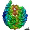
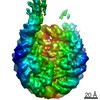
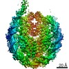
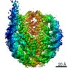
Movie
Movie Controller

[English] 日本語
 Yorodumi
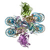
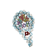
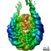
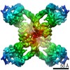
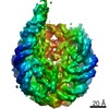
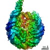
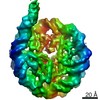
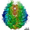
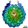
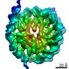
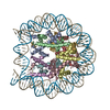
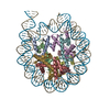
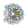
- PDB-7kbd: Nucleosome in interphase chromosome formed in Xenopus egg extract... -
+ Open data

- Basic information
| Entry | Database: PDB / ID: 7kbd | |||||||||
|---|---|---|---|---|---|---|---|---|---|---|
| Title | Nucleosome in interphase chromosome formed in Xenopus egg extract (oligo fraction) | |||||||||
 Components Components |
| |||||||||
 Keywords Keywords | STRUCTURAL PROTEIN/DNA / nucleosome / interphase / cell cycle / chromatin / DNA BINDING PROTEIN / STRUCTURAL PROTEIN-DNA complex | |||||||||
| Function / homology |  Function and homology information Function and homology informationstructural constituent of chromatin / nucleosome / nucleosome assembly / heterochromatin formation / protein heterodimerization activity / DNA binding / nucleoplasm / nucleus Similarity search - Function | |||||||||
| Biological species | ||||||||||
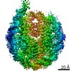
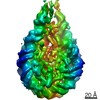
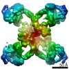
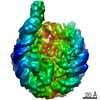
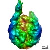
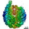
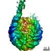
| Method | ELECTRON MICROSCOPY / single particle reconstruction / cryo EM / Resolution: 3.38 Å | |||||||||
 Authors Authors | Arimura, Y. / Funabiki, H. | |||||||||
| Funding support |  United States, United States,  Japan, 2items Japan, 2items
| |||||||||
 Citation Citation | Journal: Mol Cell / Year: 2021 Title: Structural features of nucleosomes in interphase and metaphase chromosomes. Authors: Yasuhiro Arimura / Rochelle M Shih / Ruby Froom / Hironori Funabiki / Abstract: Structural heterogeneity of nucleosomes in functional chromosomes is unknown. Here, we devise the template-, reference- and selection-free (TRSF) cryo-EM pipeline to simultaneously reconstruct cryo- ...Structural heterogeneity of nucleosomes in functional chromosomes is unknown. Here, we devise the template-, reference- and selection-free (TRSF) cryo-EM pipeline to simultaneously reconstruct cryo-EM structures of protein complexes from interphase or metaphase chromosomes. The reconstructed interphase and metaphase nucleosome structures are on average indistinguishable from canonical nucleosome structures, despite DNA sequence heterogeneity, cell-cycle-specific posttranslational modifications, and interacting proteins. Nucleosome structures determined by a decoy-classifying method and structure variability analyses reveal the nucleosome structural variations in linker DNA, histone tails, and nucleosome core particle configurations, suggesting that the opening of linker DNA, which is correlated with H2A C-terminal tail positioning, is suppressed in chromosomes. High-resolution (3.4-3.5 Å) nucleosome structures indicate DNA-sequence-independent stabilization of superhelical locations ±0-1 and ±3.5-4.5. The linker histone H1.8 preferentially binds to metaphase chromatin, from which chromatosome cryo-EM structures with H1.8 at the on-dyad position are reconstituted. This study presents the structural characteristics of nucleosomes in chromosomes. | |||||||||
| History |
|
- Structure visualization
| Movie |
Movie viewer |
|---|---|
| Structure viewer | Molecule: MolmilJmol/JSmol |
- Downloads & links
-Download
| PDBx/mmCIF format | 7kbd.cif.gz | 287.3 KB | Display | PDBx/mmCIF format |
|---|---|---|---|---|
| PDB format | pdb7kbd.ent.gz | 215.2 KB | Display | PDB format |
| PDBx/mmJSON format | 7kbd.json.gz | Tree view | PDBx/mmJSON format | |
| Others |  Other downloads Other downloads |
-Validation report
| Arichive directory | https://data.pdbj.org/pub/pdb/validation_reports/kb/7kbdftp://data.pdbj.org/pub/pdb/validation_reports/kb/7kbd | HTTPS FTP |
|---|
-Related structure data
| Related structure data | 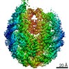 22790MC 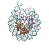 7kbeC 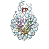 7kbfC M: map data used to model this data C: citing same article ( |
|---|---|
| Similar structure data | |
| EM raw data | EMPIAR-10692 (Title: Oligo nucleosome fraction from interphase chromosome in Xenopus egg extract lot 1 Data size: 634.3 Data #1: oligo nucleosome fraction from the interphase chromosome in Xenopus egg extract lot1 [micrographs - multiframe]) |

-Links
 PDBj
PDBj


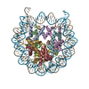
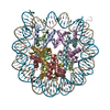
- Assembly
| Deposited unit | 
|
|---|---|
| 1 |
|
-Components
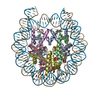
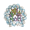
-Protein , 4 types, 8 molecules AEBFCGDH
| #1: Protein | Mass: 15421.101 Da / Num. of mol.: 2 / Source method: isolated from a natural source / Source: (natural) #2: Protein | Mass: 11394.426 Da / Num. of mol.: 2 / Source method: isolated from a natural source / Source: (natural) #3: Protein | Mass: 14883.174 Da / Num. of mol.: 2 / Source method: isolated from a natural source / Source: (natural) #4: Protein | Mass: 13965.265 Da / Num. of mol.: 2 / Source method: isolated from a natural source / Source: (natural) |
|---|
-DNA chain , 2 types, 2 molecules IJ
| #5: DNA chain | Mass: 46847.863 Da / Num. of mol.: 1 / Source method: isolated from a natural source / Source: (natural) |
|---|---|
| #6: DNA chain | Mass: 46355.543 Da / Num. of mol.: 1 / Source method: isolated from a natural source / Source: (natural) |
-Experimental details
-Experiment
| Experiment | Method: ELECTRON MICROSCOPY |
|---|---|
| EM experiment | Aggregation state: PARTICLE / 3D reconstruction method: single particle reconstruction |
- Sample preparation
| Component | Name: Nucleosome in interphase chromosome formed in Xenopus egg extract (oligo fraction) Type: COMPLEX / Entity ID: all / Source: NATURAL | |||||||||||||||||||||||||||||||||||||||||||||||||||||||
|---|---|---|---|---|---|---|---|---|---|---|---|---|---|---|---|---|---|---|---|---|---|---|---|---|---|---|---|---|---|---|---|---|---|---|---|---|---|---|---|---|---|---|---|---|---|---|---|---|---|---|---|---|---|---|---|---|
| Molecular weight | Experimental value: NO | |||||||||||||||||||||||||||||||||||||||||||||||||||||||
| Source (natural) | Organism: | |||||||||||||||||||||||||||||||||||||||||||||||||||||||
| Buffer solution | pH: 7.4 | |||||||||||||||||||||||||||||||||||||||||||||||||||||||
| Buffer component |
| |||||||||||||||||||||||||||||||||||||||||||||||||||||||
| Specimen | Embedding applied: NO / Shadowing applied: NO / Staining applied: NO / Vitrification applied: YES | |||||||||||||||||||||||||||||||||||||||||||||||||||||||
| Specimen support | Details: 20W / Grid material: GOLD / Grid mesh size: 300 divisions/in. / Grid type: Quantifoil R1.2/1.3 | |||||||||||||||||||||||||||||||||||||||||||||||||||||||
| Vitrification | Instrument: FEI VITROBOT MARK IV / Cryogen name: ETHANE / Humidity: 100 % / Chamber temperature: 277 K |
- Electron microscopy imaging
| Experimental equipment |  Model: Talos Arctica / Image courtesy: FEI Company |
|---|---|
| Microscopy | Model: FEI TALOS ARCTICA |
| Electron gun | Electron source:  FIELD EMISSION GUN / Accelerating voltage: 200 kV / Illumination mode: FLOOD BEAM FIELD EMISSION GUN / Accelerating voltage: 200 kV / Illumination mode: FLOOD BEAM |
| Electron lens | Mode: BRIGHT FIELD |
| Image recording | Electron dose: 33.11 e/Å2 / Detector mode: SUPER-RESOLUTION / Film or detector model: GATAN K2 SUMMIT (4k x 4k) |
- Processing
| Software | Name: PHENIX / Version: 1.17.1_3660: / Classification: refinement | ||||||||||||||||||||||||
|---|---|---|---|---|---|---|---|---|---|---|---|---|---|---|---|---|---|---|---|---|---|---|---|---|---|
| EM software |
| ||||||||||||||||||||||||
| CTF correction | Type: PHASE FLIPPING AND AMPLITUDE CORRECTION | ||||||||||||||||||||||||
| Symmetry | Point symmetry: C1 (asymmetric) | ||||||||||||||||||||||||
| 3D reconstruction | Resolution: 3.38 Å / Resolution method: FSC 0.143 CUT-OFF / Num. of particles: 89191 / Symmetry type: POINT | ||||||||||||||||||||||||
| Atomic model building |
| ||||||||||||||||||||||||
| Refine LS restraints |
|