 Movie
Movie Controller

[English] 日本語
 Yorodumi
- PDB-5nl0: Crystal structure of a 197-bp palindromic 601L nucleosome in comp... -
+ Open data

- Basic information
| Entry | Database: PDB / ID: 5nl0 | |||||||||||||||||||||
|---|---|---|---|---|---|---|---|---|---|---|---|---|---|---|---|---|---|---|---|---|---|---|
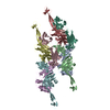
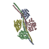
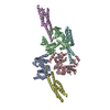
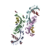
| Title | Crystal structure of a 197-bp palindromic 601L nucleosome in complex with linker histone H1 | |||||||||||||||||||||
 Components Components |
| |||||||||||||||||||||
 Keywords Keywords | CHROMATIN BINDING PROTEIN / DNA / nucleosome / chromatin / linker histones / histone H1 / CHROMATIN BINDING PROTEIN - DNA complex | |||||||||||||||||||||
| Function / homology |  Function and homology information Function and homology informationnegative regulation of DNA recombination / chromosome condensation / nucleosomal DNA binding / heterochromatin / structural constituent of chromatin / nucleosome / nucleosome assembly / heterochromatin formation / double-stranded DNA binding / protein heterodimerization activity ...negative regulation of DNA recombination / chromosome condensation / nucleosomal DNA binding / heterochromatin / structural constituent of chromatin / nucleosome / nucleosome assembly / heterochromatin formation / double-stranded DNA binding / protein heterodimerization activity / DNA binding / nucleoplasm / nucleus Similarity search - Function | |||||||||||||||||||||
| Biological species | synthetic construct (others) | |||||||||||||||||||||
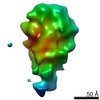
| Method |  X-RAY DIFFRACTION / SYNCHROTRON / MOLECULAR REPLACEMENT / Resolution: 5.4 Å X-RAY DIFFRACTION / SYNCHROTRON / MOLECULAR REPLACEMENT / Resolution: 5.4 Å | |||||||||||||||||||||
 Authors Authors | Garcia-Saez, I. / Petosa, C. / Dimitrov, S. | |||||||||||||||||||||
| Funding support |  France, 6items France, 6items
| |||||||||||||||||||||
 Citation Citation | Journal: Mol Cell / Year: 2017 Title: Structure and Dynamics of a 197 bp Nucleosome in Complex with Linker Histone H1. Authors: Jan Bednar / Isabel Garcia-Saez / Ramachandran Boopathi / Amber R Cutter / Gabor Papai / Anna Reymer / Sajad H Syed / Imtiaz Nisar Lone / Ognyan Tonchev / Corinne Crucifix / Hervé Menoni / ...Authors: Jan Bednar / Isabel Garcia-Saez / Ramachandran Boopathi / Amber R Cutter / Gabor Papai / Anna Reymer / Sajad H Syed / Imtiaz Nisar Lone / Ognyan Tonchev / Corinne Crucifix / Hervé Menoni / Christophe Papin / Dimitrios A Skoufias / Hitoshi Kurumizaka / Richard Lavery / Ali Hamiche / Jeffrey J Hayes / Patrick Schultz / Dimitar Angelov / Carlo Petosa / Stefan Dimitrov /   Abstract: Linker histones associate with nucleosomes to promote the formation of higher-order chromatin structure, but the underlying molecular details are unclear. We investigated the structure of a 197 bp ...Linker histones associate with nucleosomes to promote the formation of higher-order chromatin structure, but the underlying molecular details are unclear. We investigated the structure of a 197 bp nucleosome bearing symmetric 25 bp linker DNA arms in complex with vertebrate linker histone H1. We determined electron cryo-microscopy (cryo-EM) and crystal structures of unbound and H1-bound nucleosomes and validated these structures by site-directed protein cross-linking and hydroxyl radical footprinting experiments. Histone H1 shifts the conformational landscape of the nucleosome by drawing the two linkers together and reducing their flexibility. The H1 C-terminal domain (CTD) localizes primarily to a single linker, while the H1 globular domain contacts the nucleosome dyad and both linkers, associating more closely with the CTD-distal linker. These findings reveal that H1 imparts a strong degree of asymmetry to the nucleosome, which is likely to influence the assembly and architecture of higher-order structures. | |||||||||||||||||||||
| History |
|
- Structure visualization
| Structure viewer | Molecule: MolmilJmol/JSmol |
|---|
- Downloads & links
-Download
| PDBx/mmCIF format | 5nl0.cif.gz | 1.1 MB | Display | PDBx/mmCIF format |
|---|---|---|---|---|
| PDB format | pdb5nl0.ent.gz | 935.7 KB | Display | PDB format |
| PDBx/mmJSON format | 5nl0.json.gz | Tree view | PDBx/mmJSON format | |
| Others |  Other downloads Other downloads |
-Validation report
| Arichive directory | https://data.pdbj.org/pub/pdb/validation_reports/nl/5nl0ftp://data.pdbj.org/pub/pdb/validation_reports/nl/5nl0 | HTTPS FTP |
|---|
-Related structure data
| Related structure data | 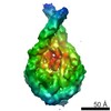 3657C 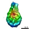 3659C 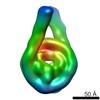 3660C 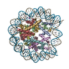 3ut9S S: Starting model for refinement C: citing same article ( |
|---|---|
| Similar structure data |

-Links
 PDBj
PDBj


- Assembly
| Deposited unit | 
| ||||||||
|---|---|---|---|---|---|---|---|---|---|
| 1 | 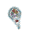
| ||||||||
| 2 | 
| ||||||||
| Unit cell |
|
-Components
-Protein , 5 types, 13 molecules AEKBFLCGMDHNZ
| #1: Protein | Mass: 15303.930 Da / Num. of mol.: 3 Source method: isolated from a genetically manipulated source Details: The core histones in the uploaded coordinates are those from X. laevis while those in the crystal are human. The reason for the discrepancy is that the structure was solved using pdb 3UT9 as ...Details: The core histones in the uploaded coordinates are those from X. laevis while those in the crystal are human. The reason for the discrepancy is that the structure was solved using pdb 3UT9 as a molecular replacement template (because it had the same 601L DNA sequence and therefore was the closest high-res structure available). Source: (gene. exp.)  #2: Protein | Mass: 11263.231 Da / Num. of mol.: 3 Source method: isolated from a genetically manipulated source Details: The core histones in the uploaded coordinates are those from X. laevis while those in the crystal are human. The reason for the discrepancy is that the structure was solved using pdb 3UT9 as ...Details: The core histones in the uploaded coordinates are those from X. laevis while those in the crystal are human. The reason for the discrepancy is that the structure was solved using pdb 3UT9 as a molecular replacement template (because it had the same 601L DNA sequence and therefore was the closest high-res structure available). Source: (gene. exp.) #3: Protein | Mass: 13978.241 Da / Num. of mol.: 3 Source method: isolated from a genetically manipulated source Details: The core histones in the uploaded coordinates are those from X. laevis while those in the crystal are human. The reason for the discrepancy is that the structure was solved using pdb 3UT9 as ...Details: The core histones in the uploaded coordinates are those from X. laevis while those in the crystal are human. The reason for the discrepancy is that the structure was solved using pdb 3UT9 as a molecular replacement template (because it had the same 601L DNA sequence and therefore was the closest high-res structure available). Source: (gene. exp.) #4: Protein | Mass: 13524.752 Da / Num. of mol.: 3 Source method: isolated from a genetically manipulated source Details: The core histones in the uploaded coordinates are those from X. laevis while those in the crystal are human. The reason for the discrepancy is that the structure was solved using pdb 3UT9 as ...Details: The core histones in the uploaded coordinates are those from X. laevis while those in the crystal are human. The reason for the discrepancy is that the structure was solved using pdb 3UT9 as a molecular replacement template (because it had the same 601L DNA sequence and therefore was the closest high-res structure available). Source: (gene. exp.) #7: Protein | | Mass: 21129.502 Da / Num. of mol.: 1 Source method: isolated from a genetically manipulated source Details: Only the H1.0b globular domain was visible in the structure. The coordinates of this chain are those of a homology model built from chicken GH5 (pdb 1HST). Source: (gene. exp.) |
|---|
-DNA chain , 2 types, 4 molecules ISJT
| #5: DNA chain | Mass: 60824.777 Da / Num. of mol.: 2 Source method: isolated from a genetically manipulated source Details: There are only 193 bp in the structure whereas there are 197 bp in the crystal; putatively this could be because two base pairs at the tip of each linker are denatured and therefore not visible. Source: (gene. exp.) synthetic construct (others) / Production host: #6: DNA chain | Mass: 60815.762 Da / Num. of mol.: 2 Source method: isolated from a genetically manipulated source Details: There are only 193 bp in the structure whereas there are 197 bp in the crystal; putatively this could be because two base pairs at the tip of each linker are denatured and therefore not visible. Source: (gene. exp.) synthetic construct (others) / Production host: |
|---|
-Experimental details
-Experiment
| Experiment | Method: X-RAY DIFFRACTION / Number of used crystals: 1 |
|---|
- Sample preparation
| Crystal | Density Matthews: 3.21 Å3/Da / Density % sol: 61.68 % / Description: Long thin rods. |
|---|---|
| Crystal grow | Temperature: 293.15 K / Method: vapor diffusion, hanging drop / pH: 6.4 Details: Mix of equal volumes of the nucleosome/H1 complex (25-30 microM) and a crystallization solution composed of MPD (6% v/v), 50 mM NaCl, and 50 mM sodium potassium phosphate pH 6.4. |
-Data collection
| Diffraction | Mean temperature: 100 K |
|---|---|
| Diffraction source | Source: SYNCHROTRON / Site: ESRF / Beamline: ID29 / Wavelength: 0.99987 Å |
| Detector | Type: DECTRIS PILATUS 6M / Detector: PIXEL / Date: Aug 29, 2015 |
| Radiation | Protocol: SINGLE WAVELENGTH / Monochromatic (M) / Laue (L): M / Scattering type: x-ray |
| Radiation wavelength | Wavelength: 0.99987 Å / Relative weight: 1 |
| Reflection | Resolution: 5.4→49.13 Å / Num. obs: 15641 / % possible obs: 99.5 % / Redundancy: 4.5 % / CC1/2: 0.997 / Rmerge(I) obs: 0.091 / Rpim(I) all: 0.072 / Net I/σ(I): 9.1 |
| Reflection shell | Resolution: 5.4→6.04 Å / Redundancy: 4.4 % / Rmerge(I) obs: 0.685 / Mean I/σ(I) obs: 2.1 / Num. unique obs: 4370 / CC1/2: 0.686 / Rpim(I) all: 0.553 / % possible all: 99.6 |
- Processing
| Software |
| ||||||||||||||||||||||||||||||||||||||||||||||||||||||||||||||||||||||||||||||||||||
|---|---|---|---|---|---|---|---|---|---|---|---|---|---|---|---|---|---|---|---|---|---|---|---|---|---|---|---|---|---|---|---|---|---|---|---|---|---|---|---|---|---|---|---|---|---|---|---|---|---|---|---|---|---|---|---|---|---|---|---|---|---|---|---|---|---|---|---|---|---|---|---|---|---|---|---|---|---|---|---|---|---|---|---|---|---|
| Refinement | Method to determine structure: MOLECULAR REPLACEMENT Starting model: 3UT9 Resolution: 5.4→49.128 Å / SU ML: 0.85 / Cross valid method: FREE R-VALUE / σ(F): 1.35 / Phase error: 31.59 Details: The asymmetric unit contains a complete H1/nucleosome complex (molecule A) and half of a second complex (molecule B). Rigid-body and TLS refinement in Phenix were performed. Linker DNA was ...Details: The asymmetric unit contains a complete H1/nucleosome complex (molecule A) and half of a second complex (molecule B). Rigid-body and TLS refinement in Phenix were performed. Linker DNA was extended from the nucleosome core as B-form DNA, adjusted into density using Coot and subjected to rigid body and TLS refinement in Phenix, resulting in improved R-values. The 2Fo-Fc and Fo-Fc maps calculated using phases from the NCP and linker DNA revealed additional density corresponding to the GH1 domain in both complexes A and B. The structure of X. laevis GH1.0b was modeled by homology with the crystal structure of chicken GH5 (PDB 1HST), which shares 80% amino acid sequence identity with X. laevis GH1.0b.
| ||||||||||||||||||||||||||||||||||||||||||||||||||||||||||||||||||||||||||||||||||||
| Solvent computation | Shrinkage radii: 0.9 Å / VDW probe radii: 1.11 Å | ||||||||||||||||||||||||||||||||||||||||||||||||||||||||||||||||||||||||||||||||||||
| Refinement step | Cycle: LAST / Resolution: 5.4→49.128 Å
| ||||||||||||||||||||||||||||||||||||||||||||||||||||||||||||||||||||||||||||||||||||
| Refine LS restraints |
| ||||||||||||||||||||||||||||||||||||||||||||||||||||||||||||||||||||||||||||||||||||
| LS refinement shell |
|
