 Movie
Movie Controller

[English] 日本語
 Yorodumi
- PDB-3ut9: Crystal Structure of Nucleosome Core Particle Assembled with a Pa... -
+ Open data

- Basic information
| Entry | Database: PDB / ID: 3ut9 | ||||||
|---|---|---|---|---|---|---|---|
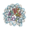
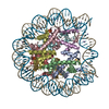
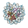
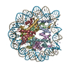
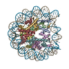
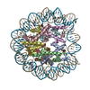
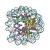
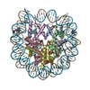
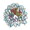
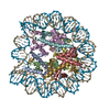
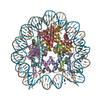
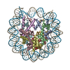
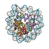
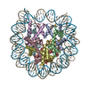
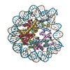
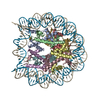
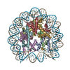
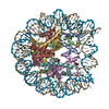
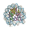
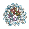
| Title | Crystal Structure of Nucleosome Core Particle Assembled with a Palindromic Widom '601' Derivative (NCP-601L) | ||||||
 Components Components |
| ||||||
 Keywords Keywords | STRUCTURAL PROTEIN/DNA / Nucleosome core particle / NCP / 601-sequence DNA / STRUCTURAL PROTEIN-DNA complex | ||||||
| Function / homology |  Function and homology information Function and homology informationstructural constituent of chromatin / nucleosome / nucleosome assembly / heterochromatin formation / protein heterodimerization activity / DNA binding / nucleoplasm / nucleus Similarity search - Function | ||||||
| Biological species | |||||||
| Method |  X-RAY DIFFRACTION / SYNCHROTRON / MOLECULAR REPLACEMENT / Resolution: 2.2 Å X-RAY DIFFRACTION / SYNCHROTRON / MOLECULAR REPLACEMENT / Resolution: 2.2 Å | ||||||
 Authors Authors | Chua, E.Y.D. / Vasudevan, D. / Davey, G.E. / Wu, B. / Davey, C.A. | ||||||
 Citation Citation | Journal: Nucleic Acids Res. / Year: 2012 Title: The mechanics behind DNA sequence-dependent properties of the nucleosome Authors: Chua, E.Y.D. / Vasudevan, D. / Davey, G.E. / Wu, B. / Davey, C.A. | ||||||
| History |
|
- Structure visualization
| Structure viewer | Molecule: MolmilJmol/JSmol |
|---|
- Downloads & links
-Download
| PDBx/mmCIF format | 3ut9.cif.gz | 328 KB | Display | PDBx/mmCIF format |
|---|---|---|---|---|
| PDB format | pdb3ut9.ent.gz | 247 KB | Display | PDB format |
| PDBx/mmJSON format | 3ut9.json.gz | Tree view | PDBx/mmJSON format | |
| Others |  Other downloads Other downloads |
-Validation report
| Arichive directory | https://data.pdbj.org/pub/pdb/validation_reports/ut/3ut9ftp://data.pdbj.org/pub/pdb/validation_reports/ut/3ut9 | HTTPS FTP |
|---|
-Related structure data


-Links
 PDBj
PDBj


- Assembly
| Deposited unit | 
| ||||||||
|---|---|---|---|---|---|---|---|---|---|
| 1 |
| ||||||||
| Unit cell |
|
-Components
-Protein , 4 types, 8 molecules AEBFCGDH
| #1: Protein | Mass: 15303.930 Da / Num. of mol.: 2 Source method: isolated from a genetically manipulated source Source: (gene. exp.)  #2: Protein | Mass: 11263.231 Da / Num. of mol.: 2 Source method: isolated from a genetically manipulated source Source: (gene. exp.) #3: Protein | Mass: 13978.241 Da / Num. of mol.: 2 Source method: isolated from a genetically manipulated source Source: (gene. exp.) #4: Protein | Mass: 13848.097 Da / Num. of mol.: 2 Source method: isolated from a genetically manipulated source Source: (gene. exp.) |
|---|
-DNA chain , 2 types, 2 molecules IJ
| #5: DNA chain | Mass: 44761.523 Da / Num. of mol.: 1 / Source method: obtained synthetically / Details: Synthetic construct |
|---|---|
| #6: DNA chain | Mass: 44752.508 Da / Num. of mol.: 1 / Source method: obtained synthetically / Details: Synthetic construct |
-Non-polymers , 4 types, 173 molecules 


| #7: Chemical |  Mass: 35.453 Da / Num. of mol.: 2 / Source method: obtained synthetically / Formula: Cl Mass: 35.453 Da / Num. of mol.: 2 / Source method: obtained synthetically / Formula: Cl#8: Chemical | ChemComp-MN /  Mass: 54.938 Da / Num. of mol.: 29 / Source method: obtained synthetically / Formula: Mn Mass: 54.938 Da / Num. of mol.: 29 / Source method: obtained synthetically / Formula: Mn#9: Chemical |  Mass: 39.098 Da / Num. of mol.: 2 / Source method: obtained synthetically / Formula: K Mass: 39.098 Da / Num. of mol.: 2 / Source method: obtained synthetically / Formula: K#10: Water | ChemComp-HOH / | Mass: 18.015 Da / Num. of mol.: 140 / Source method: isolated from a natural source / Formula: H2O |
|---|
-Details
| Sequence details | UNINTENTIO |
|---|
-Experimental details
-Experiment
| Experiment | Method: X-RAY DIFFRACTION / Number of used crystals: 1 |
|---|
- Sample preparation
| Crystal | Density Matthews: 2.57 Å3/Da / Density % sol: 52.16 % / Mosaicity: 0.3 ° |
|---|---|
| Crystal grow | Temperature: 291 K / Method: vapor diffusion, hanging drop / pH: 6 Details: K-cacodylate, KCl, MnCl2, pH 6.0, temperature 291K, VAPOR DIFFUSION, HANGING DROP |
-Data collection
| Diffraction source | Source: SYNCHROTRON / Site: SLS  / Beamline: X06SA / Wavelength: 0.8 Å / Beamline: X06SA / Wavelength: 0.8 Å | ||||||||||||||||||||||||||||||||||||||||||||||||||||||||||||||||||||||||||||||||||||||||||||||||||||||||||||||||||||||||||||||||||||
|---|---|---|---|---|---|---|---|---|---|---|---|---|---|---|---|---|---|---|---|---|---|---|---|---|---|---|---|---|---|---|---|---|---|---|---|---|---|---|---|---|---|---|---|---|---|---|---|---|---|---|---|---|---|---|---|---|---|---|---|---|---|---|---|---|---|---|---|---|---|---|---|---|---|---|---|---|---|---|---|---|---|---|---|---|---|---|---|---|---|---|---|---|---|---|---|---|---|---|---|---|---|---|---|---|---|---|---|---|---|---|---|---|---|---|---|---|---|---|---|---|---|---|---|---|---|---|---|---|---|---|---|---|---|
| Detector | Type: PSI PILATUS 6M / Detector: PIXEL | ||||||||||||||||||||||||||||||||||||||||||||||||||||||||||||||||||||||||||||||||||||||||||||||||||||||||||||||||||||||||||||||||||||
| Radiation | Protocol: SINGLE WAVELENGTH / Monochromatic (M) / Laue (L): M / Scattering type: x-ray | ||||||||||||||||||||||||||||||||||||||||||||||||||||||||||||||||||||||||||||||||||||||||||||||||||||||||||||||||||||||||||||||||||||
| Radiation wavelength | Wavelength: 0.8 Å / Relative weight: 1 | ||||||||||||||||||||||||||||||||||||||||||||||||||||||||||||||||||||||||||||||||||||||||||||||||||||||||||||||||||||||||||||||||||||
| Reflection | Resolution: 2.2→92.819 Å / Num. all: 104105 / Num. obs: 104105 / % possible obs: 99.8 % / Redundancy: 5.4 % / Rsym value: 0.054 / Net I/σ(I): 12.3 | ||||||||||||||||||||||||||||||||||||||||||||||||||||||||||||||||||||||||||||||||||||||||||||||||||||||||||||||||||||||||||||||||||||
| Reflection shell | Diffraction-ID: 1
|
- Processing
| Software |
| ||||||||||||||||||||||||||||||||||||||||||||||||||||||||||||||||||||||||||||||||||||||||||
|---|---|---|---|---|---|---|---|---|---|---|---|---|---|---|---|---|---|---|---|---|---|---|---|---|---|---|---|---|---|---|---|---|---|---|---|---|---|---|---|---|---|---|---|---|---|---|---|---|---|---|---|---|---|---|---|---|---|---|---|---|---|---|---|---|---|---|---|---|---|---|---|---|---|---|---|---|---|---|---|---|---|---|---|---|---|---|---|---|---|---|---|
| Refinement | Method to determine structure: MOLECULAR REPLACEMENT / Resolution: 2.2→42.55 Å / Cor.coef. Fo:Fc: 0.949 / Cor.coef. Fo:Fc free: 0.941 / WRfactor Rfree: 0.3263 / WRfactor Rwork: 0.2782 / Occupancy max: 1 / Occupancy min: 1 / FOM work R set: 0.7123 / SU B: 9.836 / SU ML: 0.244 / SU R Cruickshank DPI: 0.272 / SU Rfree: 0.2216 / Cross valid method: THROUGHOUT / σ(F): 0 / ESU R: 0.272 / ESU R Free: 0.222 / Stereochemistry target values: MAXIMUM LIKELIHOOD / Details: HYDROGENS HAVE BEEN ADDED IN THE RIDING POSITIONS
| ||||||||||||||||||||||||||||||||||||||||||||||||||||||||||||||||||||||||||||||||||||||||||
| Solvent computation | Ion probe radii: 0.8 Å / Shrinkage radii: 0.8 Å / VDW probe radii: 1.2 Å / Solvent model: MASK | ||||||||||||||||||||||||||||||||||||||||||||||||||||||||||||||||||||||||||||||||||||||||||
| Displacement parameters | Biso max: 199.99 Å2 / Biso mean: 87.9913 Å2 / Biso min: 12.67 Å2
| ||||||||||||||||||||||||||||||||||||||||||||||||||||||||||||||||||||||||||||||||||||||||||
| Refinement step | Cycle: LAST / Resolution: 2.2→42.55 Å
| ||||||||||||||||||||||||||||||||||||||||||||||||||||||||||||||||||||||||||||||||||||||||||
| Refine LS restraints |
| ||||||||||||||||||||||||||||||||||||||||||||||||||||||||||||||||||||||||||||||||||||||||||
| LS refinement shell | Resolution: 2.2→2.257 Å / Total num. of bins used: 20
|
