 Movie
Movie Controller

[English] 日本語
 Yorodumi
- PDB-7efw: Crystal structure of hexameric state of C-phycocyanin from Thermo... -
+ Open data

- Basic information
| Entry | Database: PDB / ID: 7efw | ||||||||||||
|---|---|---|---|---|---|---|---|---|---|---|---|---|---|
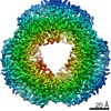
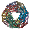
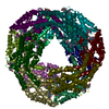
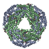
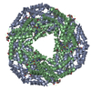
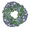
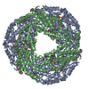
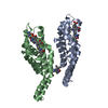
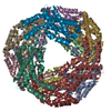
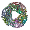
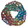
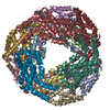
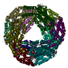
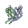
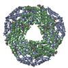
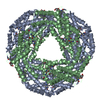
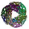
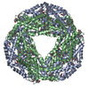
| Title | Crystal structure of hexameric state of C-phycocyanin from Thermoleptolyngbya sp. O-77 | ||||||||||||
 Components Components |
| ||||||||||||
 Keywords Keywords | PHOTOSYNTHESIS / light-harvesting complex | ||||||||||||
| Function / homology |  Function and homology information Function and homology information: / phycobilisome / plasma membrane-derived thylakoid membrane / photosynthesis Similarity search - Function | ||||||||||||
| Biological species |  Leptolyngbya sp. O-77 (bacteria) Leptolyngbya sp. O-77 (bacteria) | ||||||||||||
| Method |  X-RAY DIFFRACTION / SYNCHROTRON / MOLECULAR REPLACEMENT / Resolution: 1.65 Å X-RAY DIFFRACTION / SYNCHROTRON / MOLECULAR REPLACEMENT / Resolution: 1.65 Å | ||||||||||||
 Authors Authors | Minato, T. / Teramoto, T. / Hung, N.K. / Yamada, K. / Ogo, S. / Kakuta, Y. / Yoon, K.S. | ||||||||||||
| Funding support |  Japan, 3items Japan, 3items
| ||||||||||||
 Citation Citation | Journal: Commun Biol / Year: 2021 Title: Non-conventional octameric structure of C-phycocyanin. Authors: Takuo Minato / Takamasa Teramoto / Naruhiko Adachi / Nguyen Khac Hung / Kaho Yamada / Masato Kawasaki / Masato Akutsu / Toshio Moriya / Toshiya Senda / Seiji Ogo / Yoshimitsu Kakuta / Ki-Seok Yoon / Abstract: C-phycocyanin (CPC), a blue pigment protein, is an indispensable component of giant phycobilisomes, which are light-harvesting antenna complexes in cyanobacteria that transfer energy efficiently to ...C-phycocyanin (CPC), a blue pigment protein, is an indispensable component of giant phycobilisomes, which are light-harvesting antenna complexes in cyanobacteria that transfer energy efficiently to photosystems I and II. X-ray crystallographic and electron microscopy (EM) analyses have revealed the structure of CPC to be a closed toroidal hexamer by assembling two trimers. In this study, the structural characterization of non-conventional octameric CPC is reported for the first time. Analyses of the crystal and cryogenic EM structures of the native CPC from filamentous thermophilic cyanobacterium Thermoleptolyngbya sp. O-77 unexpectedly illustrated the coexistence of conventional hexamer and novel octamer. In addition, an unusual dimeric state, observed via analytical ultracentrifugation, was postulated to be a key intermediate structure in the assemble of the previously unobserved octamer. These observations provide new insights into the assembly processes of CPCs and the mechanism of energy transfer in the light-harvesting complexes. | ||||||||||||
| History |
|
- Structure visualization
| Structure viewer | Molecule: MolmilJmol/JSmol |
|---|
- Downloads & links
-Download
| PDBx/mmCIF format | 7efw.cif.gz | 857 KB | Display | PDBx/mmCIF format |
|---|---|---|---|---|
| PDB format | pdb7efw.ent.gz | 721.3 KB | Display | PDB format |
| PDBx/mmJSON format | 7efw.json.gz | Tree view | PDBx/mmJSON format | |
| Others |  Other downloads Other downloads |
-Validation report
| Arichive directory | https://data.pdbj.org/pub/pdb/validation_reports/ef/7efwftp://data.pdbj.org/pub/pdb/validation_reports/ef/7efw | HTTPS FTP |
|---|
-Related structure data
| Related structure data |  7efvC 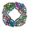 7eh7C  7eh8C 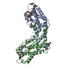 1i7yS S: Starting model for refinement C: citing same article ( |
|---|---|
| Similar structure data |


-Links
 PDBj
PDBj- Assembly
| Deposited unit | 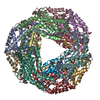
| ||||||||
|---|---|---|---|---|---|---|---|---|---|
| 1 |
| ||||||||
| Unit cell |
|
-Components
| #1: Protein | Mass: 17560.531 Da / Num. of mol.: 6 / Source method: isolated from a natural source / Source: (natural) Leptolyngbya sp. O-77 (bacteria) / References: UniProt: A0A0X8WU90#2: Protein | Mass: 18097.465 Da / Num. of mol.: 6 / Source method: isolated from a natural source / Source: (natural) Leptolyngbya sp. O-77 (bacteria) / References: UniProt: A0A0X8WUE0#3: Chemical | ChemComp-CYC /   Mass: 588.694 Da / Num. of mol.: 18 / Source method: obtained synthetically / Formula: C33H40N4O6 / Feature type: SUBJECT OF INVESTIGATION Mass: 588.694 Da / Num. of mol.: 18 / Source method: obtained synthetically / Formula: C33H40N4O6 / Feature type: SUBJECT OF INVESTIGATION#4: Chemical |   Mass: 92.094 Da / Num. of mol.: 3 / Source method: obtained synthetically / Formula: C3H8O3 Mass: 92.094 Da / Num. of mol.: 3 / Source method: obtained synthetically / Formula: C3H8O3#5: Water | ChemComp-HOH / |  Mass: 18.015 Da / Num. of mol.: 2676 / Source method: isolated from a natural source / Formula: H2O Mass: 18.015 Da / Num. of mol.: 2676 / Source method: isolated from a natural source / Formula: H2OHas ligand of interest | Y | |
|---|
-Experimental details
-Experiment
| Experiment | Method: X-RAY DIFFRACTION / Number of used crystals: 1 |
|---|
- Sample preparation
| Crystal | Density Matthews: 2.76 Å3/Da / Density % sol: 55.51 % |
|---|---|
| Crystal grow | Temperature: 293.15 K / Method: vapor diffusion, sitting drop / pH: 5 Details: 8% Tacsimate pH 5.0, 20 % Polyethylene glycol (PEG) 3350 |
-Data collection
| Diffraction | Mean temperature: 100 K / Serial crystal experiment: N |
|---|---|
| Diffraction source | Source: SYNCHROTRON / Site: SPring-8 / Beamline: BL45XU / Wavelength: 1 Å |
| Detector | Type: DECTRIS PILATUS3 6M / Detector: PIXEL / Date: Oct 17, 2020 |
| Radiation | Protocol: SINGLE WAVELENGTH / Monochromatic (M) / Laue (L): M / Scattering type: x-ray |
| Radiation wavelength | Wavelength: 1 Å / Relative weight: 1 |
| Reflection | Resolution: 1.65→49.2 Å / Num. obs: 264074 / % possible obs: 92.4 % / Redundancy: 5.9 % / CC1/2: 0.993 / Rmerge(I) obs: 0.194 / Rpim(I) all: 0.083 / Rrim(I) all: 0.221 / Net I/σ(I): 8.6 |
| Reflection shell | Resolution: 1.65→1.74 Å / Redundancy: 2.9 % / Num. unique obs: 25606 / CC1/2: 0.495 / Rrim(I) all: 1.062 / % possible all: 65.1 |
- Processing
| Software |
| |||||||||||||||||||||||||||||||||||||||||||||||||||||||||||||||||||||||||||||||||||||||||||||||||||||||||
|---|---|---|---|---|---|---|---|---|---|---|---|---|---|---|---|---|---|---|---|---|---|---|---|---|---|---|---|---|---|---|---|---|---|---|---|---|---|---|---|---|---|---|---|---|---|---|---|---|---|---|---|---|---|---|---|---|---|---|---|---|---|---|---|---|---|---|---|---|---|---|---|---|---|---|---|---|---|---|---|---|---|---|---|---|---|---|---|---|---|---|---|---|---|---|---|---|---|---|---|---|---|---|---|---|---|---|
| Refinement | Method to determine structure: MOLECULAR REPLACEMENT Starting model: 1I7Y Resolution: 1.65→49.18 Å / SU ML: 0.14 / Cross valid method: FREE R-VALUE / σ(F): 1.36 / Phase error: 19.13 / Stereochemistry target values: ML
| |||||||||||||||||||||||||||||||||||||||||||||||||||||||||||||||||||||||||||||||||||||||||||||||||||||||||
| Solvent computation | Shrinkage radii: 0.9 Å / VDW probe radii: 1.11 Å / Solvent model: FLAT BULK SOLVENT MODEL | |||||||||||||||||||||||||||||||||||||||||||||||||||||||||||||||||||||||||||||||||||||||||||||||||||||||||
| Refinement step | Cycle: LAST / Resolution: 1.65→49.18 Å
| |||||||||||||||||||||||||||||||||||||||||||||||||||||||||||||||||||||||||||||||||||||||||||||||||||||||||
| Refine LS restraints |
| |||||||||||||||||||||||||||||||||||||||||||||||||||||||||||||||||||||||||||||||||||||||||||||||||||||||||
| LS refinement shell |
| |||||||||||||||||||||||||||||||||||||||||||||||||||||||||||||||||||||||||||||||||||||||||||||||||||||||||
| Refinement TLS params. | Method: refined / Origin x: -0.1873 Å / Origin y: -45.0847 Å / Origin z: 21.8752 Å
| |||||||||||||||||||||||||||||||||||||||||||||||||||||||||||||||||||||||||||||||||||||||||||||||||||||||||
| Refinement TLS group | Selection details: all |
