 Movie
Movie Controller

[English] 日本語
 Yorodumi
- PDB-7eh7: Cryo-EM structure of the octameric state of C-phycocyanin from Th... -
+ Open data

- Basic information
| Entry | Database: PDB / ID: 7eh7 | |||||||||||||||
|---|---|---|---|---|---|---|---|---|---|---|---|---|---|---|---|---|
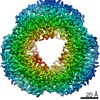
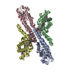
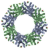
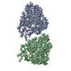
| Title | Cryo-EM structure of the octameric state of C-phycocyanin from Thermoleptolyngbya sp. O-77 | |||||||||||||||
 Components Components |
| |||||||||||||||
 Keywords Keywords | PHOTOSYNTHESIS / homo hexamer / light-harvesting complex | |||||||||||||||
| Function / homology |  Function and homology information Function and homology information: / phycobilisome / plasma membrane-derived thylakoid membrane / photosynthesis Similarity search - Function | |||||||||||||||
| Biological species |  Leptolyngbya sp. O-77 (bacteria) Leptolyngbya sp. O-77 (bacteria) | |||||||||||||||
| Method | ELECTRON MICROSCOPY / single particle reconstruction / cryo EM / Resolution: 3.71 Å | |||||||||||||||
 Authors Authors | Minato, T. / Teramoto, T. / Adachi, N. / Hung, N.K. / Yamada, K. / Kawasaki, M. / Akutsu, M. / Moriya, T. / Senda, T. / Ogo, S. ...Minato, T. / Teramoto, T. / Adachi, N. / Hung, N.K. / Yamada, K. / Kawasaki, M. / Akutsu, M. / Moriya, T. / Senda, T. / Ogo, S. / Kakuta, Y. / Yoon, K.S. | |||||||||||||||
| Funding support |  Japan, 4items Japan, 4items
| |||||||||||||||
 Citation Citation | Journal: Commun Biol / Year: 2021 Title: Non-conventional octameric structure of C-phycocyanin. Authors: Takuo Minato / Takamasa Teramoto / Naruhiko Adachi / Nguyen Khac Hung / Kaho Yamada / Masato Kawasaki / Masato Akutsu / Toshio Moriya / Toshiya Senda / Seiji Ogo / Yoshimitsu Kakuta / Ki-Seok Yoon / Abstract: C-phycocyanin (CPC), a blue pigment protein, is an indispensable component of giant phycobilisomes, which are light-harvesting antenna complexes in cyanobacteria that transfer energy efficiently to ...C-phycocyanin (CPC), a blue pigment protein, is an indispensable component of giant phycobilisomes, which are light-harvesting antenna complexes in cyanobacteria that transfer energy efficiently to photosystems I and II. X-ray crystallographic and electron microscopy (EM) analyses have revealed the structure of CPC to be a closed toroidal hexamer by assembling two trimers. In this study, the structural characterization of non-conventional octameric CPC is reported for the first time. Analyses of the crystal and cryogenic EM structures of the native CPC from filamentous thermophilic cyanobacterium Thermoleptolyngbya sp. O-77 unexpectedly illustrated the coexistence of conventional hexamer and novel octamer. In addition, an unusual dimeric state, observed via analytical ultracentrifugation, was postulated to be a key intermediate structure in the assemble of the previously unobserved octamer. These observations provide new insights into the assembly processes of CPCs and the mechanism of energy transfer in the light-harvesting complexes. | |||||||||||||||
| History |
|
- Structure visualization
| Movie |
Movie viewer |
|---|---|
| Structure viewer | Molecule: MolmilJmol/JSmol |
- Downloads & links
-Download
| PDBx/mmCIF format | 7eh7.cif.gz | 442.8 KB | Display | PDBx/mmCIF format |
|---|---|---|---|---|
| PDB format | pdb7eh7.ent.gz | 379.3 KB | Display | PDB format |
| PDBx/mmJSON format | 7eh7.json.gz | Tree view | PDBx/mmJSON format | |
| Others |  Other downloads Other downloads |
-Validation report
| Arichive directory | https://data.pdbj.org/pub/pdb/validation_reports/eh/7eh7ftp://data.pdbj.org/pub/pdb/validation_reports/eh/7eh7 | HTTPS FTP |
|---|
-Related structure data
| Related structure data |  31089MC  7efvC 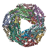 7efwC  7eh8C M: map data used to model this data C: citing same article ( |
|---|---|
| Similar structure data | |
| EM raw data | EMPIAR-11079 (Title: Non-conventional octameric structure of C-phycocyanin Data size: 1.8 TB Data #1: Non-conventional octameric structure of C-phycocyanin [micrographs - multiframe]) |


-Links
 PDBj
PDBj- Assembly
| Deposited unit | 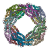
|
|---|---|
| 1 |
|
-Components
| #1: Protein | Mass: 17560.531 Da / Num. of mol.: 8 / Source method: isolated from a natural source / Source: (natural) Leptolyngbya sp. O-77 (bacteria) / References: UniProt: A0A0X8WU90#2: Protein | Mass: 18097.465 Da / Num. of mol.: 8 / Source method: isolated from a natural source / Details: MEN: N-METHYL ASPARAGINE / Source: (natural) Leptolyngbya sp. O-77 (bacteria) / References: UniProt: A0A0X8WUE0#3: Chemical | ChemComp-CYC /   Mass: 588.694 Da / Num. of mol.: 24 / Source method: obtained synthetically / Formula: C33H40N4O6 / Feature type: SUBJECT OF INVESTIGATION Mass: 588.694 Da / Num. of mol.: 24 / Source method: obtained synthetically / Formula: C33H40N4O6 / Feature type: SUBJECT OF INVESTIGATIONHas ligand of interest | Y | Has protein modification | Y | |
|---|
-Experimental details
-Experiment
| Experiment | Method: ELECTRON MICROSCOPY |
|---|---|
| EM experiment | Aggregation state: PARTICLE / 3D reconstruction method: single particle reconstruction |
- Sample preparation
| Component | Name: C-phycocyanin octamer / Type: COMPLEX / Entity ID: #1-#2 / Source: NATURAL |
|---|---|
| Molecular weight | Value: 0.29 MDa / Experimental value: NO |
| Source (natural) | Organism: Leptolyngbya sp. O-77 (bacteria) |
| Buffer solution | pH: 7 |
| Specimen | Conc.: 6.7 mg/ml / Embedding applied: NO / Shadowing applied: NO / Staining applied: NO / Vitrification applied: YES / Details: This sample was mono-disperse. |
| Specimen support | Details: The grid was washed by acetone prior to use. / Grid material: COPPER / Grid mesh size: 300 divisions/in. / Grid type: Quantifoil R1.2/1.3 |
| Vitrification | Instrument: FEI VITROBOT MARK IV / Cryogen name: ETHANE / Humidity: 100 % / Chamber temperature: 291 K / Details: Blotting time was 5 seconds (blot force 15) |
- Electron microscopy imaging
| Microscopy | Model: TFS TALOS |
|---|---|
| Electron gun | Electron source:  FIELD EMISSION GUN / Accelerating voltage: 200 kV / Illumination mode: FLOOD BEAM FIELD EMISSION GUN / Accelerating voltage: 200 kV / Illumination mode: FLOOD BEAM |
| Electron lens | Mode: BRIGHT FIELD / Nominal magnification: 120000 X / Nominal defocus max: 2500 nm / Nominal defocus min: 1000 nm / Cs: 2.7 mm / C2 aperture diameter: 50 µm |
| Specimen holder | Cryogen: NITROGEN |
| Image recording | Average exposure time: 49.2 sec. / Electron dose: 50 e/Å2 / Detector mode: COUNTING / Film or detector model: FEI FALCON III (4k x 4k) / Num. of grids imaged: 2 / Num. of real images: 2036 |
- Processing
| Software | Name: PHENIX / Version: 1.19_4092: / Classification: refinement | ||||||||||||||||||||||||||||||||
|---|---|---|---|---|---|---|---|---|---|---|---|---|---|---|---|---|---|---|---|---|---|---|---|---|---|---|---|---|---|---|---|---|---|
| EM software |
| ||||||||||||||||||||||||||||||||
| CTF correction | Type: PHASE FLIPPING AND AMPLITUDE CORRECTION | ||||||||||||||||||||||||||||||||
| Particle selection | Num. of particles selected: 129653 | ||||||||||||||||||||||||||||||||
| Symmetry | Point symmetry: D4 (2x4 fold dihedral) | ||||||||||||||||||||||||||||||||
| 3D reconstruction | Resolution: 3.71 Å / Resolution method: FSC 0.143 CUT-OFF / Num. of particles: 10402 / Algorithm: FOURIER SPACE / Num. of class averages: 1 / Symmetry type: POINT | ||||||||||||||||||||||||||||||||
| Refine LS restraints |
|
