 Movie
Movie Controller

[English] 日本語
 Yorodumi
- PDB-3flt: Crystal structure of PE-bound octameric SAP-like pentraxin from L... -
+ Open data

- Basic information
| Entry | Database: PDB / ID: 3flt | ||||||
|---|---|---|---|---|---|---|---|
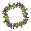
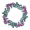
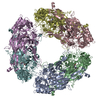
| Title | Crystal structure of PE-bound octameric SAP-like pentraxin from Limulus polyphemus | ||||||
 Components Components | SAP-like pentraxin | ||||||
 Keywords Keywords | SUGAR BINDING PROTEIN / PENTRAXIN FOLD / PHYSIOLOGICAL DOUBLY-STACKED OCTAMER / CYCLIC OCTAMER / INVERTEBRATE LECTIN | ||||||
| Function / homology |  Function and homology information Function and homology information | ||||||
| Biological species |  Limulus polyphemus (Atlantic horseshoe crab) Limulus polyphemus (Atlantic horseshoe crab) | ||||||
| Method |  X-RAY DIFFRACTION / SYNCHROTRON / Resolution: 2.7 Å X-RAY DIFFRACTION / SYNCHROTRON / Resolution: 2.7 Å | ||||||
 Authors Authors | Shrive, A.K. / Greenhough, T.J. / Armstrong, P.B. | ||||||
 Citation Citation | Journal: J Mol Biol / Year: 2009 Title: Crystal structures of Limulus SAP-like pentraxin reveal two molecular aggregations. Authors: Annette K Shrive / Ian Burns / Hui-Ting Chou / Henning Stahlberg / Peter B Armstrong / Trevor J Greenhough /  Abstract: The serum-amyloid-P-component-like pentraxin from Limulus polyphemus, a recently discovered pentraxin species and important effector protein of the hemolymph immune system, displays two distinct ...The serum-amyloid-P-component-like pentraxin from Limulus polyphemus, a recently discovered pentraxin species and important effector protein of the hemolymph immune system, displays two distinct doubly stacked cyclic molecular aggregations, heptameric and octameric. The refined three-dimensional structures determined by X-ray crystallography, both based on the same cDNA sequence, show that each aggregate is constructed from a similar dimer of protomers, which is repeated to make up the ring structure. The native octameric form has been refined at a resolution of 3 A, the native heptameric form at 2.3 A, and the phosphoethanolamine (PE)-bound octameric form at 2.7 A. The existence of the hitherto undescribed heptameric form was confirmed by single-particle analysis using cryo-electron microscopy. In the native structures, the calcium-binding site is similar to that in human pentraxins, with two calcium ions bound in each subunit. Upon binding PE, however, each subunit binds a third calcium ion, with all three calcium ions contributing to the binding and orientation of the bound phosphate group within the ligand-binding pocket. While the phosphate is well-defined in the electron density, the ethanolamine group is poorly defined, suggesting structural and binding variabilities of this group. Although sequence homology with human serum amyloid P component is relatively low, structural homology is high, with very similar overall folds and a common affinity for PE. This is due, in part, to a "topological" equivalence of side-chain position. Identical side chains that are important in both function and fold, from different regions of the sequence in human and Limulus structures, occupy similar space within the overall subunit fold. Sequence and structure alignment, based on the refined three-dimensional structures presented here and the known horseshoe crab pentraxin sequences, suggest that adaptation and refinement of C-reactive-protein-mediated immune responses in these ancient creatures lacking antibody-based immunity are based on adaptation by gene duplication. #1: Journal: J.Mol.Biol. / Year: 1999Title: C-reactive protein and SAP-like pentraxin are both present in Limulus polyphemus haemolymph: Crystal structure of Limulus SAP Authors: Shrive, A.K. / Metcalfe, A.M. / Cartwright, J.R. / Greenhough, T.J. #2: Journal: J.Mol.Biol. / Year: 2002 Title: Complete cDNA sequence of SAP-like pentraxin from Limulus polyphemus: Implications for pentraxin evolution Authors: Tharia, H.A. / Shrive, A.K. / Mills, J.D. / Arme, C. / Williams, G.T. / Greenhough, T.J. | ||||||
| History |
|
- Structure visualization
| Structure viewer | Molecule: MolmilJmol/JSmol |
|---|
- Downloads & links
-Download
| PDBx/mmCIF format | 3flt.cif.gz | 96.6 KB | Display | PDBx/mmCIF format |
|---|---|---|---|---|
| PDB format | pdb3flt.ent.gz | 73.9 KB | Display | PDB format |
| PDBx/mmJSON format | 3flt.json.gz | Tree view | PDBx/mmJSON format | |
| Others |  Other downloads Other downloads |
-Validation report
| Arichive directory | https://data.pdbj.org/pub/pdb/validation_reports/fl/3fltftp://data.pdbj.org/pub/pdb/validation_reports/fl/3flt | HTTPS FTP |
|---|
-Related structure data
| Related structure data |  1598C  3flpC  3flrSC S: Starting model for refinement C: citing same article ( |
|---|---|
| Similar structure data |


-Links
 PDBj
PDBj
- Assembly
| Deposited unit | 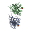
| ||||||||
|---|---|---|---|---|---|---|---|---|---|
| 1 | x 8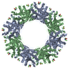
| ||||||||
| Unit cell |
| ||||||||
| Details | The biological assembly is a doubly-stacked octamer generated from the dimer in the asymmetric unit (Chains A and B) |
-Components
| #1: Protein | Mass: 23808.756 Da / Num. of mol.: 2 / Source method: isolated from a natural source / Details: Haemolymph Source: (natural) Limulus polyphemus (Atlantic horseshoe crab)References: UniProt: Q8WQK3 #2: Chemical | ChemComp-CA /   Mass: 40.078 Da / Num. of mol.: 6 / Source method: obtained synthetically / Formula: Ca Mass: 40.078 Da / Num. of mol.: 6 / Source method: obtained synthetically / Formula: Ca#3: Chemical | 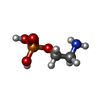  Mass: 141.063 Da / Num. of mol.: 2 / Source method: obtained synthetically / Formula: C2H8NO4P Mass: 141.063 Da / Num. of mol.: 2 / Source method: obtained synthetically / Formula: C2H8NO4P |
|---|
-Experimental details
-Experiment
| Experiment | Method: X-RAY DIFFRACTION / Number of used crystals: 1 |
|---|
- Sample preparation
| Crystal | Density Matthews: 3.84 Å3/Da / Density % sol: 67.94 % |
|---|---|
| Crystal grow | Temperature: 298 K / Method: vapor diffusion, sitting drop / pH: 7 Details: PEG 6000, CaCl2, MES, PE, pH 7, VAPOR DIFFUSION, SITTING DROP, temperature 298.0K |
-Data collection
| Diffraction | Mean temperature: 100 K |
|---|---|
| Diffraction source | Source: SYNCHROTRON / Site: SRS / Beamline: PX14.1 / Wavelength: 1.488 Å |
| Detector | Type: ADSC QUANTUM 4 / Detector: CCD / Date: Apr 3, 2005 |
| Radiation | Protocol: SINGLE WAVELENGTH / Scattering type: x-ray |
| Radiation wavelength | Wavelength: 1.488 Å / Relative weight: 1 |
| Reflection | Resolution: 2.7→60.681 Å / Num. all: 20530 / Num. obs: 20530 / % possible obs: 99.6 % / Redundancy: 3.8 % / Rmerge(I) obs: 0.084 / Rsym value: 0.084 / Net I/σ(I): 4.975 |
| Reflection shell | Resolution: 2.7→2.85 Å / Redundancy: 3.9 % / Rmerge(I) obs: 0.252 / Mean I/σ(I) obs: 2.6 / Num. measured all: 11379 / Num. unique all: 2928 / Rsym value: 0.252 / % possible all: 98.9 |
- Processing
| Software |
| |||||||||||||||||||||||||
|---|---|---|---|---|---|---|---|---|---|---|---|---|---|---|---|---|---|---|---|---|---|---|---|---|---|---|
| Refinement | Starting model: Crystals were isomorphous to octameric native. Native structure, pdb code 3FLR, was used in rigid body refinement. Resolution: 2.7→60.68 Å / Occupancy max: 1 / Occupancy min: 1 / Isotropic thermal model: Isotropic / Cross valid method: THROUGHOUT / σ(F): 0 / Stereochemistry target values: Engh & Huber
| |||||||||||||||||||||||||
| Solvent computation | Bsol: 20.309 Å2 | |||||||||||||||||||||||||
| Displacement parameters | Biso max: 59.73 Å2 / Biso mean: 28.337 Å2 / Biso min: 10.4 Å2
| |||||||||||||||||||||||||
| Refinement step | Cycle: LAST / Resolution: 2.7→60.68 Å
| |||||||||||||||||||||||||
| Refine LS restraints |
| |||||||||||||||||||||||||
| LS refinement shell | Resolution: 2.7→2.8 Å
| |||||||||||||||||||||||||
| Xplor file |
|
