 Movie
Movie Controller

[English] 日本語
 Yorodumi
- PDB-1v4l: Crystal structure of a platelet agglutination factor isolated fro... -
+ Open data

- Basic information
| Entry | Database: PDB / ID: 1v4l | ||||||
|---|---|---|---|---|---|---|---|
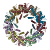
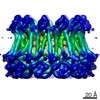
| Title | Crystal structure of a platelet agglutination factor isolated from the venom of Taiwan habu (Trimeresurus mucrosquamatus) | ||||||
 Components Components |
| ||||||
 Keywords Keywords | BLOOD CLOTTING / lectin-like / square-shaped ring | ||||||
| Function / homology |  Function and homology information Function and homology information | ||||||
| Biological species |  Protobothrops mucrosquamatus (brown spotted pit viper) Protobothrops mucrosquamatus (brown spotted pit viper) | ||||||
| Method |  X-RAY DIFFRACTION / SYNCHROTRON / MOLECULAR REPLACEMENT / Resolution: 2.8 Å X-RAY DIFFRACTION / SYNCHROTRON / MOLECULAR REPLACEMENT / Resolution: 2.8 Å | ||||||
 Authors Authors | Huang, K.-F. / Ko, T.-P. / Wang, A.H.-J. | ||||||
 Citation Citation | Journal: Biochem.J. / Year: 2004 Title: Crystal structure of a platelet-agglutinating factor isolated from the venom of Taiwan habu (Trimeresurus mucrosquamatus). Authors: Huang, K.F. / Ko, T.P. / Hung, C.C. / Chu, J. / Wang, A.H. / Chiou, S.H. | ||||||
| History |
|
- Structure visualization
| Structure viewer | Molecule: MolmilJmol/JSmol |
|---|
- Downloads & links
-Download
| PDBx/mmCIF format | 1v4l.cif.gz | 178 KB | Display | PDBx/mmCIF format |
|---|---|---|---|---|
| PDB format | pdb1v4l.ent.gz | 142.4 KB | Display | PDB format |
| PDBx/mmJSON format | 1v4l.json.gz | Tree view | PDBx/mmJSON format | |
| Others |  Other downloads Other downloads |
-Validation report
| Arichive directory | https://data.pdbj.org/pub/pdb/validation_reports/v4/1v4lftp://data.pdbj.org/pub/pdb/validation_reports/v4/1v4l | HTTPS FTP |
|---|
-Related structure data
| Related structure data |  1c3aS S: Starting model for refinement |
|---|---|
| Similar structure data |


-Links
 PDBj
PDBj


- Assembly
| Deposited unit | 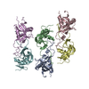
| ||||||||||||||||||||||||
|---|---|---|---|---|---|---|---|---|---|---|---|---|---|---|---|---|---|---|---|---|---|---|---|---|---|
| 1 | 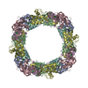
| ||||||||||||||||||||||||
| 2 | 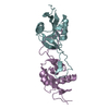
| ||||||||||||||||||||||||
| 3 | 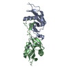
| ||||||||||||||||||||||||
| 4 | 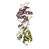
| ||||||||||||||||||||||||
| 5 | 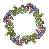
| ||||||||||||||||||||||||
| 6 | x 8
| ||||||||||||||||||||||||
| Unit cell |
| ||||||||||||||||||||||||
| Components on special symmetry positions |
| ||||||||||||||||||||||||
| Details | The biological assembly is a tetramer generated from the dimer in the asymmetric unit |
-Components
| #1: Protein | Mass: 15745.479 Da / Num. of mol.: 3 / Source method: isolated from a natural source Source: (natural) Protobothrops mucrosquamatus (brown spotted pit viper)Secretion: snake venom / References: UniProt: Q6TPH0 #2: Protein | Mass: 14559.232 Da / Num. of mol.: 3 / Source method: isolated from a natural source Source: (natural) Protobothrops mucrosquamatus (brown spotted pit viper)Secretion: snake venom / References: UniProt: Q6TPG9 #3: Water | ChemComp-HOH / |  Mass: 18.015 Da / Num. of mol.: 536 / Source method: isolated from a natural source / Formula: H2O Mass: 18.015 Da / Num. of mol.: 536 / Source method: isolated from a natural source / Formula: H2OHas protein modification | Y | |
|---|
-Experimental details
-Experiment
| Experiment | Method: X-RAY DIFFRACTION / Number of used crystals: 1 |
|---|
- Sample preparation
| Crystal | Density Matthews: 3.32 Å3/Da / Density % sol: 62.64 % | ||||||||||||||||||||||||
|---|---|---|---|---|---|---|---|---|---|---|---|---|---|---|---|---|---|---|---|---|---|---|---|---|---|
| Crystal grow | Temperature: 298 K / Method: vapor diffusion, hanging drop / pH: 5.6 Details: 1,6-hexanediol, sodium citrate, pH 5.6, VAPOR DIFFUSION, HANGING DROP, temperature 298K | ||||||||||||||||||||||||
| Crystal grow | *PLUS Method: vapor diffusion, hanging drop | ||||||||||||||||||||||||
| Components of the solutions | *PLUS
|
-Data collection
| Diffraction | Mean temperature: 100 K |
|---|---|
| Diffraction source | Source: SYNCHROTRON / Site: NSRRC  / Beamline: BL17B2 / Wavelength: 1 Å / Beamline: BL17B2 / Wavelength: 1 Å |
| Detector | Type: RIGAKU RAXIS IV / Detector: IMAGE PLATE / Date: Mar 25, 2003 |
| Radiation | Protocol: SINGLE WAVELENGTH / Monochromatic (M) / Laue (L): M / Scattering type: x-ray |
| Radiation wavelength | Wavelength: 1 Å / Relative weight: 1 |
| Reflection | Resolution: 2.8→20 Å / Num. all: 32858 / Num. obs: 30509 / % possible obs: 92.9 % / Observed criterion σ(F): 0 / Observed criterion σ(I): 0 / Redundancy: 4.2 % / Rmerge(I) obs: 0.1 / Net I/σ(I): 11.5 |
| Reflection shell | Resolution: 2.8→2.9 Å / Redundancy: 4.2 % / Rmerge(I) obs: 0.482 / Mean I/σ(I) obs: 2.9 / Num. unique all: 2971 / % possible all: 92.3 |
| Reflection | *PLUS Lowest resolution: 20 Å / Num. measured all: 127650 / Rmerge(I) obs: 0.1 |
| Reflection shell | *PLUS % possible obs: 92.33 % / Num. unique obs: 2971 / Num. measured obs: 12356 |
- Processing
| Software |
| |||||||||||||||||||||||||
|---|---|---|---|---|---|---|---|---|---|---|---|---|---|---|---|---|---|---|---|---|---|---|---|---|---|---|
| Refinement | Method to determine structure: MOLECULAR REPLACEMENT Starting model: PDB ENTRY 1C3A Resolution: 2.8→18 Å / Cross valid method: THROUGHOUT / σ(F): 0 / σ(I): 0 / Stereochemistry target values: Engh & Huber
| |||||||||||||||||||||||||
| Refine analyze | Luzzati coordinate error obs: 0.42 Å / Luzzati sigma a obs: 0.57 Å | |||||||||||||||||||||||||
| Refinement step | Cycle: LAST / Resolution: 2.8→18 Å
| |||||||||||||||||||||||||
| Refine LS restraints |
| |||||||||||||||||||||||||
| LS refinement shell | Resolution: 2.8→2.9 Å
| |||||||||||||||||||||||||
| Refinement | *PLUS Lowest resolution: 18 Å / % reflection Rfree: 5 % / Rfactor Rfree: 0.294 | |||||||||||||||||||||||||
| Solvent computation | *PLUS | |||||||||||||||||||||||||
| Displacement parameters | *PLUS |
