 Movie
Movie Controller


+ Open data

- Basic information
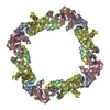
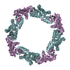
| Entry | Database: PDB / ID: 1qtj | ||||||
|---|---|---|---|---|---|---|---|
| Title | CRYSTAL STRUCTURE OF LIMULUS POLYPHEMUS SAP | ||||||
 Components Components | PROTEIN (SERUM AMYLOID P COMPONENT) | ||||||
 Keywords Keywords | SUGAR BINDING PROTEIN / PENTRAXIN FOLD / PHYSIOLOGICAL DOUBLY-STACKED OCTAMER / CYCLIC OCTAMER / INVERTEBRATE LECTIN | ||||||
| Biological species |  Limulus polyphemus (Atlantic horseshoe crab) Limulus polyphemus (Atlantic horseshoe crab) | ||||||
| Method |  X-RAY DIFFRACTION / SYNCHROTRON / MOLECULAR REPLACEMENT / Resolution: 3 Å X-RAY DIFFRACTION / SYNCHROTRON / MOLECULAR REPLACEMENT / Resolution: 3 Å | ||||||
 Authors Authors | Shrive, A.K. / Metcalfe, A.M. / Cartwright, J.R. / Greenhough, T.J. | ||||||
 Citation Citation | Journal: J.Mol.Biol. / Year: 1999 Title: C-reactive protein and SAP-like pentraxin are both present in Limulus polyphemus haemolymph: crystal structure of Limulus SAP. Authors: Shrive, A.K. / Metcalfe, A.M. / Cartwright, J.R. / Greenhough, T.J. #1: Journal: Nat.Struct.Biol. / Year: 1996Title: Three Dimensional Structure of Human C-reactive Protein Authors: Shrive, A.K. / Cheetham, G.M.T. / Holden, D. / Myles, D.A.A. / Turnell, W.G. / Volanakis, J.E. / Pepys, M.B. / Bloomer, A.C. / Greenhough, T.J. #2: Journal: J.Mol.Biol. / Year: 1990Title: Preliminary Crystallographic Analysis of C-reactive Protein from Limulus Polyphemus Authors: Myles, D.A.A. / Bailey, S. / Rule, S.A. / Jones, G.R. / Greenhough, T.J. #3: Journal: J.Biol.Chem. / Year: 1996Title: A Cytolytic Function for a Sialic Acid-Binding Lectin That is a Member of the Pentraxin Family of Proteins Authors: Armstrong, P.B. / Swarnakar, S. / Srimal, S. / Misquith, S. / Hahn, E.A. / Aimes, R.T. / Quigley, J.P. | ||||||
| History |
|
- Structure visualization
| Structure viewer | Molecule:  MolmilJmol/JSmol MolmilJmol/JSmol |
|---|
- Downloads & links
-Download
| PDBx/mmCIF format | 1qtj.cif.gz | 51.1 KB | Display | PDBx/mmCIF format |
|---|---|---|---|---|
| PDB format | pdb1qtj.ent.gz | 40.2 KB | Display | PDB format |
| PDBx/mmJSON format | 1qtj.json.gz | Tree view | PDBx/mmJSON format | |
| Others |  Other downloads Other downloads |
-Validation report
| Arichive directory | https://data.pdbj.org/pub/pdb/validation_reports/qt/1qtjftp://data.pdbj.org/pub/pdb/validation_reports/qt/1qtj | HTTPS FTP |
|---|
-Related structure data
| Related structure data | 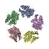 1gnhS S: Starting model for refinement |
|---|---|
| Similar structure data |

-Links
 PDBj
PDBj- Assembly
| Deposited unit | 
| |||||||||
|---|---|---|---|---|---|---|---|---|---|---|
| 1 | x 8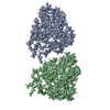
| |||||||||
| Unit cell |
| |||||||||
| Noncrystallographic symmetry (NCS) | NCS domain:
| |||||||||
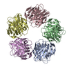
| Details | The biological assembly is a hexadecamer constructed of two cyclic octamers. This is generated by application of space group symmetry to the dimer ( protomers A and B) in the crystallographic asymmetric unit. THE BIOLOGICAL ASSEMBLY IS A HEXADECAMER CONSTRUCTED OF TWO CYCLIC OCTAMERS. |
-Components
| #1: Protein | Mass: 18485.723 Da / Num. of mol.: 2 / Source method: isolated from a natural source Source: (natural) Limulus polyphemus (Atlantic horseshoe crab)Tissue: HAEMOLYMPH |
|---|
-Experimental details
-Experiment
| Experiment | Method: X-RAY DIFFRACTION / Number of used crystals: 10 |
|---|
- Sample preparation
| Crystal | Density Matthews: 3.85 Å3/Da / Density % sol: 68 % | ||||||||||||||||||||||||
|---|---|---|---|---|---|---|---|---|---|---|---|---|---|---|---|---|---|---|---|---|---|---|---|---|---|
| Crystal grow | *PLUS pH: 7 / Method: vapor diffusion, hanging drop | ||||||||||||||||||||||||
| Components of the solutions | *PLUS
|
-Data collection
| Diffraction | Mean temperature: 298 K |
|---|---|
| Diffraction source | Source: SYNCHROTRON / Site: SRS  / Beamline: PX7.2 / Wavelength: 1.488 / Beamline: PX7.2 / Wavelength: 1.488 |
| Detector | Type: CEA / Detector: FILM / Date: Jan 1, 1989 |
| Radiation | Protocol: SINGLE WAVELENGTH / Monochromatic (M) / Laue (L): M / Scattering type: x-ray |
| Radiation wavelength | Wavelength: 1.488 Å / Relative weight: 1 |
| Reflection | Resolution: 3→60 Å / Num. all: 13666 / Num. obs: 13666 / % possible obs: 89 % / Observed criterion σ(F): 0 / Observed criterion σ(I): 0 / Redundancy: 4 % / Rmerge(I) obs: 0.117 |
| Reflection shell | Resolution: 3→3.13 Å / Rmerge(I) obs: 0.284 / % possible all: 55.6 |
| Reflection | *PLUS Num. measured all: 47412 |
| Reflection shell | *PLUS % possible obs: 55.6 % |
- Processing
| Software |
| ||||||||||||||||||||||||||||||
|---|---|---|---|---|---|---|---|---|---|---|---|---|---|---|---|---|---|---|---|---|---|---|---|---|---|---|---|---|---|---|---|
| Refinement | Method to determine structure: MOLECULAR REPLACEMENT Starting model: HUMAN C-REACTIVE PROTEIN PROTOMER (1GNH) Resolution: 3→15 Å / Data cutoff high absF: 1000000 / Data cutoff low absF: 0.001 / σ(F): 0 Details: The polyalanine structure is a final fine-tune O fitting of the X-PLOR refined polyalanine chains A and B to the subsequently modified and averaged difference density. The NCS correlation in ...Details: The polyalanine structure is a final fine-tune O fitting of the X-PLOR refined polyalanine chains A and B to the subsequently modified and averaged difference density. The NCS correlation in DM between the two independent protomers was 0.938.
| ||||||||||||||||||||||||||||||
| Refinement step | Cycle: LAST / Resolution: 3→15 Å
| ||||||||||||||||||||||||||||||
| Refine LS restraints NCS |
| ||||||||||||||||||||||||||||||
| Software | *PLUS Name: X-PLOR / Version: 3.851 / Classification: refinement | ||||||||||||||||||||||||||||||
| Refine LS restraints | *PLUS
|
