 Movie
Movie Controller

[English] 日本語
 Yorodumi
- PDB-1umr: Crystal structure of the platelet activator convulxin, a disulfid... -
+ Open data

- Basic information
| Entry | Database: PDB / ID: 1umr | ||||||
|---|---|---|---|---|---|---|---|
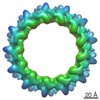
| Title | Crystal structure of the platelet activator convulxin, a disulfide linked a4b4 cyclic tetramer from the venom of Crotalus durissus terrificus | ||||||
 Components Components |
| ||||||
 Keywords Keywords | SUGAR BINDING PROTEIN / LECTIN / C-TYPE LECTIN / PLATELET / SUGAR-BINDING PROTEIN / ACTIVATOR / SNAKE VENOM | ||||||
| Function / homology |  Function and homology information Function and homology informationvenom-mediated suppression of platelet aggregation / modulation of process of another organism / positive regulation of platelet aggregation / toxin activity / extracellular region Similarity search - Function | ||||||
| Biological species |  CROTALUS DURISSUS TERRIFICUS (tropical rattlesnake) CROTALUS DURISSUS TERRIFICUS (tropical rattlesnake) | ||||||
| Method |  X-RAY DIFFRACTION / MOLECULAR REPLACEMENT / Resolution: 2.4 Å X-RAY DIFFRACTION / MOLECULAR REPLACEMENT / Resolution: 2.4 Å | ||||||
 Authors Authors | Murakami, M.T. / Zela, S.P. / Gava, L.M. / Michelan-Duarte, S. / Cintra, A.C.O. / Arni, R.K. | ||||||
 Citation Citation | Journal: Biochem.Biophys.Res.Commun. / Year: 2003 Title: Crystal Structure of the Platelet Activator Convulxin, a Disulfide Linked A4B4 Cyclic Tetramer from the Venom of Crotalus Durissus Terrificus Authors: Murakami, M.T. / Zela, S.P. / Gava, L.M. / Michelan-Duarte, S. / Cintra, A.C.O. / Arni, R.K. #1: Journal: Acta Crystallogr.,Sect.D / Year: 2003 Title: Initial Structural Analysis of an Alpha(4)Beta(4) C-Type Lectin from the Venom of Crotalus Durissus Terrificus Authors: Murakami, M.T. / Watanabe, L. / Gava, L.M. / Zela, S.P. / Cintra, A.C.O. / Arni, R.K. | ||||||
| History |
| ||||||
| Remark 700 | SHEET THE SHEET STRUCTURE OF THIS MOLECULE IS BIFURCATED. IN ORDER TO REPRESENT THIS FEATURE IN ... SHEET THE SHEET STRUCTURE OF THIS MOLECULE IS BIFURCATED. IN ORDER TO REPRESENT THIS FEATURE IN THE SHEET RECORDS BELOW, TWO SHEETS ARE DEFINED. |
- Structure visualization
| Structure viewer | Molecule: MolmilJmol/JSmol |
|---|
- Downloads & links
-Download
| PDBx/mmCIF format | 1umr.cif.gz | 122.8 KB | Display | PDBx/mmCIF format |
|---|---|---|---|---|
| PDB format | pdb1umr.ent.gz | 97.1 KB | Display | PDB format |
| PDBx/mmJSON format | 1umr.json.gz | Tree view | PDBx/mmJSON format | |
| Others |  Other downloads Other downloads |
-Validation report
| Arichive directory | https://data.pdbj.org/pub/pdb/validation_reports/um/1umrftp://data.pdbj.org/pub/pdb/validation_reports/um/1umr | HTTPS FTP |
|---|
-Related structure data
| Related structure data |  1c3aS S: Starting model for refinement |
|---|---|
| Similar structure data |
-Links
 PDBj
PDBj


- Assembly
| Deposited unit | 
| ||||||||
|---|---|---|---|---|---|---|---|---|---|
| 1 | 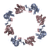
| ||||||||
| 2 | 
| ||||||||
| 3 | 
| ||||||||
| Unit cell |
| ||||||||
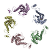
| Details | THE QUATERNARY STRUCTURE IS OF THE TYPE A4B4 CYCLICTETRAMER (HETERO-OCTAMERIC) |
-Components
| #1: Protein | Mass: 15745.936 Da / Num. of mol.: 2 / Source method: isolated from a natural source Source: (natural) CROTALUS DURISSUS TERRIFICUS (tropical rattlesnake)Tissue: VENOM GLAND / References: UniProt: O93426 #2: Protein | Mass: 14960.623 Da / Num. of mol.: 2 / Source method: isolated from a natural source Source: (natural) CROTALUS DURISSUS TERRIFICUS (tropical rattlesnake)Tissue: VENOM GLAND / References: UniProt: O93427 #3: Water | ChemComp-HOH / |  Mass: 18.015 Da / Num. of mol.: 197 / Source method: isolated from a natural source / Formula: H2O Mass: 18.015 Da / Num. of mol.: 197 / Source method: isolated from a natural source / Formula: H2OHas protein modification | Y | |
|---|
-Experimental details
-Experiment
| Experiment | Method: X-RAY DIFFRACTION / Number of used crystals: 1 |
|---|
- Sample preparation
| Crystal | Density Matthews: 4.8 Å3/Da / Density % sol: 73 % | |||||||||||||||||||||||||||||||||||
|---|---|---|---|---|---|---|---|---|---|---|---|---|---|---|---|---|---|---|---|---|---|---|---|---|---|---|---|---|---|---|---|---|---|---|---|---|
| Crystal grow | pH: 4.6 / Details: pH 4.60 | |||||||||||||||||||||||||||||||||||
| Crystal grow | *PLUS pH: 4.6 / Method: vapor diffusion | |||||||||||||||||||||||||||||||||||
| Components of the solutions | *PLUS
|
-Data collection
| Diffraction | Mean temperature: 100 K |
|---|---|
| Diffraction source | Source: ROTATING ANODE / Type: RIGAKU RU300 / Wavelength: 1.5418 |
| Detector | Type: MARRESEARCH / Detector: IMAGE PLATE / Details: MIRRORS |
| Radiation | Protocol: SINGLE WAVELENGTH / Monochromatic (M) / Laue (L): M / Scattering type: x-ray |
| Radiation wavelength | Wavelength: 1.5418 Å / Relative weight: 1 |
| Reflection | Resolution: 2.4→30 Å / Num. obs: 36900 / % possible obs: 97.6 % / Observed criterion σ(I): 2 / Redundancy: 2.4 % / Rmerge(I) obs: 0.061 / Net I/σ(I): 12.1 |
| Reflection shell | Resolution: 2.4→2.6 Å / Rmerge(I) obs: 0.404 / Mean I/σ(I) obs: 1.9 / % possible all: 98.8 |
| Reflection | *PLUS Highest resolution: 2.4 Å / Lowest resolution: 30 Å / Rmerge(I) obs: 0.061 |
| Reflection shell | *PLUS % possible obs: 98.8 % / Rmerge(I) obs: 0.404 / Mean I/σ(I) obs: 1.9 |
- Processing
| Software |
| ||||||||||||||||||||||||||||||||||||||||||||||||||||||||||||||||||||||||||||||||||||||||||||||||||||||||||||||||||||||||||||||||||||||||||||||||||||||||||||||||||||||||||||||||||||||
|---|---|---|---|---|---|---|---|---|---|---|---|---|---|---|---|---|---|---|---|---|---|---|---|---|---|---|---|---|---|---|---|---|---|---|---|---|---|---|---|---|---|---|---|---|---|---|---|---|---|---|---|---|---|---|---|---|---|---|---|---|---|---|---|---|---|---|---|---|---|---|---|---|---|---|---|---|---|---|---|---|---|---|---|---|---|---|---|---|---|---|---|---|---|---|---|---|---|---|---|---|---|---|---|---|---|---|---|---|---|---|---|---|---|---|---|---|---|---|---|---|---|---|---|---|---|---|---|---|---|---|---|---|---|---|---|---|---|---|---|---|---|---|---|---|---|---|---|---|---|---|---|---|---|---|---|---|---|---|---|---|---|---|---|---|---|---|---|---|---|---|---|---|---|---|---|---|---|---|---|---|---|---|---|
| Refinement | Method to determine structure: MOLECULAR REPLACEMENT Starting model: PDB ENTRY 1C3A Resolution: 2.4→30 Å / Cor.coef. Fo:Fc: 0.951 / Cor.coef. Fo:Fc free: 0.906 / SU B: 6.669 / SU ML: 0.155 / Cross valid method: THROUGHOUT / ESU R: 0.237 / ESU R Free: 0.234 / Stereochemistry target values: MAXIMUM LIKELIHOOD
| ||||||||||||||||||||||||||||||||||||||||||||||||||||||||||||||||||||||||||||||||||||||||||||||||||||||||||||||||||||||||||||||||||||||||||||||||||||||||||||||||||||||||||||||||||||||
| Solvent computation | Ion probe radii: 0.8 Å / Shrinkage radii: 0.8 Å / VDW probe radii: 1.4 Å / Solvent model: BABINET MODEL WITH MASK | ||||||||||||||||||||||||||||||||||||||||||||||||||||||||||||||||||||||||||||||||||||||||||||||||||||||||||||||||||||||||||||||||||||||||||||||||||||||||||||||||||||||||||||||||||||||
| Displacement parameters | Biso mean: 47.25 Å2
| ||||||||||||||||||||||||||||||||||||||||||||||||||||||||||||||||||||||||||||||||||||||||||||||||||||||||||||||||||||||||||||||||||||||||||||||||||||||||||||||||||||||||||||||||||||||
| Refinement step | Cycle: LAST / Resolution: 2.4→30 Å
| ||||||||||||||||||||||||||||||||||||||||||||||||||||||||||||||||||||||||||||||||||||||||||||||||||||||||||||||||||||||||||||||||||||||||||||||||||||||||||||||||||||||||||||||||||||||
| Refine LS restraints |
|