: / Pentaxin family / Pentraxin / C-reactive protein / pentaxin family / Pentraxin-related / Pentraxin (PTX) domain profile. / Concanavalin A-like lectin/glucanase domain superfamily / extracellular region / metal ion binding / Pentraxin family member
Function and homology information
Biological species
Limulus polyphemus (Atlantic horseshoe crab)
Method
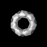
single particle reconstruction / cryo EM / negative staining / Resolution: 14.0 Å
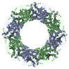
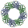
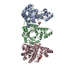
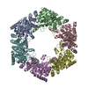
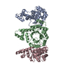
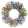
Journal: J Mol Biol / Year: 2009 Title: Crystal structures of Limulus SAP-like pentraxin reveal two molecular aggregations. Authors: Annette K Shrive / Ian Burns / Hui-Ting Chou / Henning Stahlberg / Peter B Armstrong / Trevor J Greenhough / Abstract: The serum-amyloid-P-component-like pentraxin from Limulus polyphemus, a recently discovered pentraxin species and important effector protein of the hemolymph immune system, displays two distinct ...The serum-amyloid-P-component-like pentraxin from Limulus polyphemus, a recently discovered pentraxin species and important effector protein of the hemolymph immune system, displays two distinct doubly stacked cyclic molecular aggregations, heptameric and octameric. The refined three-dimensional structures determined by X-ray crystallography, both based on the same cDNA sequence, show that each aggregate is constructed from a similar dimer of protomers, which is repeated to make up the ring structure. The native octameric form has been refined at a resolution of 3 A, the native heptameric form at 2.3 A, and the phosphoethanolamine (PE)-bound octameric form at 2.7 A. The existence of the hitherto undescribed heptameric form was confirmed by single-particle analysis using cryo-electron microscopy. In the native structures, the calcium-binding site is similar to that in human pentraxins, with two calcium ions bound in each subunit. Upon binding PE, however, each subunit binds a third calcium ion, with all three calcium ions contributing to the binding and orientation of the bound phosphate group within the ligand-binding pocket. While the phosphate is well-defined in the electron density, the ethanolamine group is poorly defined, suggesting structural and binding variabilities of this group. Although sequence homology with human serum amyloid P component is relatively low, structural homology is high, with very similar overall folds and a common affinity for PE. This is due, in part, to a "topological" equivalence of side-chain position. Identical side chains that are important in both function and fold, from different regions of the sequence in human and Limulus structures, occupy similar space within the overall subunit fold. Sequence and structure alignment, based on the refined three-dimensional structures presented here and the known horseshoe crab pentraxin sequences, suggest that adaptation and refinement of C-reactive-protein-mediated immune responses in these ancient creatures lacking antibody-based immunity are based on adaptation by gene duplication.
History
Deposition
Feb 1, 2009
-
Header (metadata) release
Feb 6, 2009
-
Map release
Apr 1, 2009
-
Update
Oct 24, 2012
-
Current status
Oct 24, 2012
Processing site: PDBe / Status: Released
-
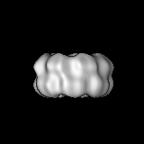
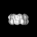
Structure visualization
Movie
Surface view with section colored by density value
Type: NEGATIVE Details: 3ul protein sample laied on the grid for 30 seconds and the grid floated on saturated ammonium molybdate for 1 minute
Grid
Details: 400 mesh quantifoil grid
Vitrification
Cryogen name: ETHANE / Chamber temperature: 100 K / Instrument: OTHER Method: The grid was blotted for 6 seconds and air-dried for 3 seconds before plunging
-
Electron microscopy
Microscope
JEOL 2100F
Temperature
Average: 95 K
Details
Imaged in JEOL minimum dose system
Image recording
Category: FILM / Film or detector model: KODAK SO-163 FILM / Digitization - Scanner: PRIMESCAN / Digitization - Sampling interval: 10 µm / Number real images: 18 / Bits/pixel: 16
Electron beam
Acceleration voltage: 120 kV / Electron source: LAB6
In the structure databanks used in Yorodumi, some data are registered as the other names, "COVID-19 virus" and "2019-nCoV". Here are the details of the virus and the list of structure data.
Jan 31, 2019. EMDB accession codes are about to change! (news from PDBe EMDB page)
EMDB accession codes are about to change! (news from PDBe EMDB page)
The allocation of 4 digits for EMDB accession codes will soon come to an end. Whilst these codes will remain in use, new EMDB accession codes will include an additional digit and will expand incrementally as the available range of codes is exhausted. The current 4-digit format prefixed with “EMD-” (i.e. EMD-XXXX) will advance to a 5-digit format (i.e. EMD-XXXXX), and so on. It is currently estimated that the 4-digit codes will be depleted around Spring 2019, at which point the 5-digit format will come into force.
The EM Navigator/Yorodumi systems omit the EMD- prefix.
Related info.:Q: What is EMD? / ID/Accession-code notation in Yorodumi/EM Navigator
Yorodumi is a browser for structure data from EMDB, PDB, SASBDB, etc.
This page is also the successor to EM Navigator detail page, and also detail information page/front-end page for Omokage search.
The word "yorodu" (or yorozu) is an old Japanese word meaning "ten thousand". "mi" (miru) is to see.
Related info.:EMDB / PDB / SASBDB / Comparison of 3 databanks / Yorodumi Search / Aug 31, 2016. New EM Navigator & Yorodumi / Yorodumi Papers / Jmol/JSmol / Function and homology information / Changes in new EM Navigator and Yorodumi
 Movie
Movie Controller

 Yorodumi
 Open data
 Basic information
 Map data
 Sample
 Keywords
 Function and homology information
 Limulus polyphemus (Atlantic horseshoe crab)
 Authors
 Citation
 Structure visualization
 UCSF Chimera
 Downloads & links
 1598.gif
 http://ftp.pdbj.org/pub/emdb/structures/EMD-1598

 Z (Sec.)
 Y (Row.)
 X (Col.)


 Sample components
 Processing
 Electron microscopy