 Movie
Movie Controller

[English] 日本語
 Yorodumi
- PDB-4fir: Crystal structure of pyridoxal biosynthesis lyase PdxS from Pyrococcus -
+ Open data

- Basic information
| Entry | Database: PDB / ID: 4fir | ||||||
|---|---|---|---|---|---|---|---|
| Title | Crystal structure of pyridoxal biosynthesis lyase PdxS from Pyrococcus | ||||||
 Components Components | Pyridoxal biosynthesis lyase pdxS | ||||||
 Keywords Keywords | LYASE / Pyrococcus horikoshii / pdxS / pyridoxal biosynthesis lyase / pyridoxal 5 -phosphate (PLP) | ||||||
| Function / homology |  Function and homology information Function and homology informationpyridoxal 5'-phosphate synthase (glutamine hydrolysing) / pyridoxal 5'-phosphate synthase (glutamine hydrolysing) activity / pyridoxal 5'-phosphate biosynthetic process / pyridoxine biosynthetic process / amino acid metabolic process Similarity search - Function | ||||||
| Biological species |   Pyrococcus horikoshii (archaea) Pyrococcus horikoshii (archaea) | ||||||
| Method |  X-RAY DIFFRACTION / SYNCHROTRON / MOLECULAR REPLACEMENT / Resolution: 3.1 Å X-RAY DIFFRACTION / SYNCHROTRON / MOLECULAR REPLACEMENT / Resolution: 3.1 Å | ||||||
 Authors Authors | Matsuura, A. / Yoon, J.Y. / Yoon, H.J. / Lee, H.H. / Suh, S.W. | ||||||
 Citation Citation | Journal: Mol.Cells / Year: 2012 Title: Crystal structure of pyridoxal biosynthesis lyase PdxS from Pyrococcus horikoshii. Authors: Matsuura, A. / Yoon, J.Y. / Yoon, H.J. / Lee, H.H. / Suh, S.W. | ||||||
| History |
|
- Structure visualization
| Structure viewer | Molecule: MolmilJmol/JSmol |
|---|
- Downloads & links
-Download
| PDBx/mmCIF format | 4fir.cif.gz | 377.6 KB | Display | PDBx/mmCIF format |
|---|---|---|---|---|
| PDB format | pdb4fir.ent.gz | 309.4 KB | Display | PDB format |
| PDBx/mmJSON format | 4fir.json.gz | Tree view | PDBx/mmJSON format | |
| Others |  Other downloads Other downloads |
-Validation report
| Arichive directory | https://data.pdbj.org/pub/pdb/validation_reports/fi/4firftp://data.pdbj.org/pub/pdb/validation_reports/fi/4fir | HTTPS FTP |
|---|
-Related structure data
| Related structure data |  4fiqC  1znnS C: citing same article ( S: Starting model for refinement |
|---|---|
| Similar structure data |
-Links
 PDBj
PDBj
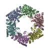
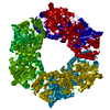
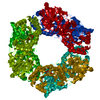
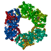
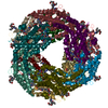
- Assembly
| Deposited unit | 
| ||||||||||||||||||||||||||||||||||||||||||
|---|---|---|---|---|---|---|---|---|---|---|---|---|---|---|---|---|---|---|---|---|---|---|---|---|---|---|---|---|---|---|---|---|---|---|---|---|---|---|---|---|---|---|---|
| 1 |
| ||||||||||||||||||||||||||||||||||||||||||
| Unit cell |
| ||||||||||||||||||||||||||||||||||||||||||
| Noncrystallographic symmetry (NCS) | NCS domain:
NCS domain segments:
|
-Components
| #1: Protein | Mass: 37065.309 Da / Num. of mol.: 6 Source method: isolated from a genetically manipulated source Source: (gene. exp.) Pyrococcus horikoshii (archaea)Strain: ATCC 700860 / DSM 12428 / JCM 9974 / NBRC 100139 / OT-3 Gene: pdxS, PH1355 / Plasmid: pET-21a(+) / Production host:  #2: Sugar | ChemComp-R5P /   Type: saccharide / Mass: 230.110 Da / Num. of mol.: 6 Type: saccharide / Mass: 230.110 Da / Num. of mol.: 6Source method: isolated from a genetically manipulated source Formula: C5H11O8P #3: Water | ChemComp-HOH / |  Mass: 18.015 Da / Num. of mol.: 72 / Source method: isolated from a natural source / Formula: H2O Mass: 18.015 Da / Num. of mol.: 72 / Source method: isolated from a natural source / Formula: H2OHas protein modification | Y | |
|---|
-Experimental details
-Experiment
| Experiment | Method: X-RAY DIFFRACTION / Number of used crystals: 1 |
|---|
- Sample preparation
| Crystal | Density Matthews: 2.54 Å3/Da / Density % sol: 51.6 % |
|---|---|
| Crystal grow | Temperature: 297 K / pH: 8 Details: 100mM imidazole, 35% 2-methyl-2,4-pentanediol, 200mM MgCl2, pH 8.0, VAPOR DIFFUSION, HANGING DROP, temperature 297K |
-Data collection
| Diffraction | Mean temperature: 100 K |
|---|---|
| Diffraction source | Source: SYNCHROTRON / Site: Photon Factory  / Beamline: BL-5A / Wavelength: 1 / Beamline: BL-5A / Wavelength: 1 |
| Detector | Type: ADSC QUANTUM 210r / Detector: CCD / Date: Nov 23, 2005 |
| Radiation | Monochromator: SI 111 CHANNEL / Protocol: SINGLE WAVELENGTH / Monochromatic (M) / Laue (L): M / Scattering type: x-ray |
| Radiation wavelength | Wavelength: 1 Å / Relative weight: 1 |
| Reflection | Resolution: 3.1→30 Å / Num. obs: 39532 / % possible obs: 98.5 % / Biso Wilson estimate: 57.7 Å2 / Rmerge(I) obs: 0.066 |
| Reflection shell | Resolution: 3.1→3.15 Å / Rmerge(I) obs: 0.23 / % possible all: 85.5 |
- Processing
| Software |
| |||||||||||||||||||||||||||||||||||||||||||||||||||||||||||||||||||||||||||||||||||||||||||||||||||||||||
|---|---|---|---|---|---|---|---|---|---|---|---|---|---|---|---|---|---|---|---|---|---|---|---|---|---|---|---|---|---|---|---|---|---|---|---|---|---|---|---|---|---|---|---|---|---|---|---|---|---|---|---|---|---|---|---|---|---|---|---|---|---|---|---|---|---|---|---|---|---|---|---|---|---|---|---|---|---|---|---|---|---|---|---|---|---|---|---|---|---|---|---|---|---|---|---|---|---|---|---|---|---|---|---|---|---|---|
| Refinement | Method to determine structure: MOLECULAR REPLACEMENT Starting model: 1ZNN Resolution: 3.1→29.84 Å / Occupancy max: 1 / Occupancy min: 1 / SU ML: 0.38 / σ(F): 1.38 / Phase error: 23.22 / Stereochemistry target values: ML
| |||||||||||||||||||||||||||||||||||||||||||||||||||||||||||||||||||||||||||||||||||||||||||||||||||||||||
| Solvent computation | Shrinkage radii: 0.86 Å / VDW probe radii: 1.1 Å / Solvent model: FLAT BULK SOLVENT MODEL / Bsol: 33.27 Å2 / ksol: 0.31 e/Å3 | |||||||||||||||||||||||||||||||||||||||||||||||||||||||||||||||||||||||||||||||||||||||||||||||||||||||||
| Displacement parameters | Biso mean: 54.21 Å2
| |||||||||||||||||||||||||||||||||||||||||||||||||||||||||||||||||||||||||||||||||||||||||||||||||||||||||
| Refinement step | Cycle: LAST / Resolution: 3.1→29.84 Å
| |||||||||||||||||||||||||||||||||||||||||||||||||||||||||||||||||||||||||||||||||||||||||||||||||||||||||
| Refine LS restraints |
| |||||||||||||||||||||||||||||||||||||||||||||||||||||||||||||||||||||||||||||||||||||||||||||||||||||||||
| Refine LS restraints NCS |
| |||||||||||||||||||||||||||||||||||||||||||||||||||||||||||||||||||||||||||||||||||||||||||||||||||||||||
| LS refinement shell |
|
