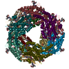
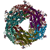
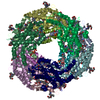
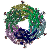
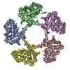
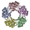
Journal: Structure / Year: 1998 Title: Three-dimensional reconstructions from cryoelectron microscopy images reveal an intimate complex between helicase DnaB and its loading partner DnaC. Authors: C San Martin / M Radermacher / B Wolpensinger / A Engel / C S Miles / N E Dixon / J M Carazo / Abstract: BACKGROUND: DNA helicases play a fundamental role in all aspects of nucleic acid metabolism and defects in these enzymes have been implicated in a number of inherited human disorders. DnaB is the ...BACKGROUND: DNA helicases play a fundamental role in all aspects of nucleic acid metabolism and defects in these enzymes have been implicated in a number of inherited human disorders. DnaB is the major replicative DNA helicase in Escherichia coli and has been used as a model system for studying the structure and function of hexameric helicases. The native protein is a hexamer of identical subunits, which in solution forms a complex with six molecules of the loading protein DnaC. DnaB is delivered from this complex onto the DNA template, with the subsequent release of DnaC. We report here the structures of the DnaB helicase hexamer and its complex with DnaC under a defined set of experimental conditions, as determined by three-dimensional cryoelectron microscopy. It was hoped that the structures would provide insight into the mechanisms of helicase activity. RESULTS: The DnaB structure reveals that six DnaB monomers assemble as three asymmetric dimers to form a polar, ring-like hexamer. The hexamer has two faces, one displaying threefold and the other ...RESULTS: The DnaB structure reveals that six DnaB monomers assemble as three asymmetric dimers to form a polar, ring-like hexamer. The hexamer has two faces, one displaying threefold and the other sixfold symmetry. The six DnaC protomers bind tightly to the sixfold face of the DnaB hexamer. This is the first report of a three-dimensional structure of a helicase obtained using cryoelectron microscopy, and the first report of the structure of a helicase in complex with a loading protein. CONCLUSIONS: The structures of the DnaB helicase and its complex with DnaC reveal some interesting structural features relevant to helicase function and to the assembly of the two-protein complex. ...CONCLUSIONS: The structures of the DnaB helicase and its complex with DnaC reveal some interesting structural features relevant to helicase function and to the assembly of the two-protein complex. The results presented here provide a basis for a more complete understanding of the structure and function of these important proteins.
History
Deposition
Feb 10, 2003
-
Header (metadata) release
Feb 12, 2003
-
Map release
Feb 12, 2003
-
Update
May 26, 2011
-
Current status
May 26, 2011
Processing site: PDBe / Status: Released
-
Structure visualization
Movie
Surface view with section colored by density value
GO: DNA helicase activity / InterPro: INTERPRO: IPR001198
-
Experimental details
-
Structure determination
Method
cryo EM
Processing
single particle reconstruction
Aggregation state
particle
-
Sample preparation
Concentration
.04 mg/mL
Buffer
pH: 7.6 Details: 50 mM Tris.HCl pH 7.6 2 mM DTT 5 mM MgCl2 200 mM NaCl 0.25 mM ADP
Vitrification
Cryogen name: ETHANE / Instrument: HOMEMADE PLUNGER Details: Vitrification instrument: double side blotting device Method: samples were adsorbed onto carbon-coated molybdenum holey grids after 30s glow discharge, and vitrified by quick plunging in liquid ethane in a double-side blotting device
-
Electron microscopy
Microscope
FEI/PHILIPS EM420
Temperature
Average: 98 K
Image recording
Category: FILM / Film or detector model: KODAK SO-163 FILM / Digitization - Scanner: EIKONIX IEEE 488 / Number real images: 27 / Average electron dose: 10 e/Å2 / Bits/pixel: 8
Electron beam
Acceleration voltage: 100 kV / Electron source: LAB6
In the structure databanks used in Yorodumi, some data are registered as the other names, "COVID-19 virus" and "2019-nCoV". Here are the details of the virus and the list of structure data.
Jan 31, 2019. EMDB accession codes are about to change! (news from PDBe EMDB page)
EMDB accession codes are about to change! (news from PDBe EMDB page)
The allocation of 4 digits for EMDB accession codes will soon come to an end. Whilst these codes will remain in use, new EMDB accession codes will include an additional digit and will expand incrementally as the available range of codes is exhausted. The current 4-digit format prefixed with “EMD-” (i.e. EMD-XXXX) will advance to a 5-digit format (i.e. EMD-XXXXX), and so on. It is currently estimated that the 4-digit codes will be depleted around Spring 2019, at which point the 5-digit format will come into force.
The EM Navigator/Yorodumi systems omit the EMD- prefix.
Related info.:Q: What is EMD? / ID/Accession-code notation in Yorodumi/EM Navigator
Yorodumi is a browser for structure data from EMDB, PDB, SASBDB, etc.
This page is also the successor to EM Navigator detail page, and also detail information page/front-end page for Omokage search.
The word "yorodu" (or yorozu) is an old Japanese word meaning "ten thousand". "mi" (miru) is to see.
Related info.:EMDB / PDB / SASBDB / Comparison of 3 databanks / Yorodumi Search / Aug 31, 2016. New EM Navigator & Yorodumi / Yorodumi Papers / Jmol/JSmol / Function and homology information / Changes in new EM Navigator and Yorodumi
 Movie
Movie Controller

 Yorodumi
 Open data
 Basic information
 Map data
 Sample
 Function and homology information
 Authors
 Citation
 Structure visualization
 UCSF Chimera
 Downloads & links
 1022.gif
 http://ftp.pdbj.org/pub/emdb/structures/EMD-1022
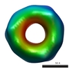


 Z (Sec.)
 Y (Row.)
 X (Col.)


 Sample components
 Processing
 Electron microscopy