[English] 日本語
Yorodumi
- PDB-3flp: Crystal structure of native heptameric SAP-like pentraxin from Li... -

+
Open data


ID or keywords:

Loading...

-
Basic information

Entry
Database: PDB / ID: 3flp
TitleCrystal structure of native heptameric SAP-like pentraxin from Limulus polyphemus
ComponentsSAP-like pentraxin
KeywordsSUGAR BINDING PROTEIN / physiological doubly-stacked heptamer / pentraxin fold / cyclic heptamer / invertebrate lectin
Function / homology
Function and homology information


extracellular region / metal ion binding
Similarity search - Function
Pentaxin family / Pentraxin / C-reactive protein / pentaxin family / Pentraxin-related / Pentraxin (PTX) domain profile. / Jelly Rolls - #200 / Concanavalin A-like lectin/glucanase domain superfamily / Jelly Rolls / Sandwich / Mainly Beta
Similarity search - Domain/homology
Pentraxin family member
Similarity search - Component
Biological speciesLimulus polyphemus (Atlantic horseshoe crab)
MethodX-RAY DIFFRACTION / SYNCHROTRON / MOLECULAR REPLACEMENT / molecular replacement / Resolution: 2.3 Å
AuthorsShrive, A.K. / Greenhough, T.J. / Armstrong, P.B.
Citation
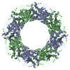
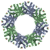
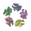
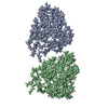
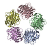
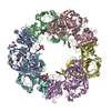
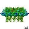
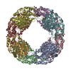
Journal: J Mol Biol / Year: 2009
Title: Crystal structures of Limulus SAP-like pentraxin reveal two molecular aggregations.
Authors: Annette K Shrive / Ian Burns / Hui-Ting Chou / Henning Stahlberg / Peter B Armstrong / Trevor J Greenhough /
Abstract: The serum-amyloid-P-component-like pentraxin from Limulus polyphemus, a recently discovered pentraxin species and important effector protein of the hemolymph immune system, displays two distinct ...The serum-amyloid-P-component-like pentraxin from Limulus polyphemus, a recently discovered pentraxin species and important effector protein of the hemolymph immune system, displays two distinct doubly stacked cyclic molecular aggregations, heptameric and octameric. The refined three-dimensional structures determined by X-ray crystallography, both based on the same cDNA sequence, show that each aggregate is constructed from a similar dimer of protomers, which is repeated to make up the ring structure. The native octameric form has been refined at a resolution of 3 A, the native heptameric form at 2.3 A, and the phosphoethanolamine (PE)-bound octameric form at 2.7 A. The existence of the hitherto undescribed heptameric form was confirmed by single-particle analysis using cryo-electron microscopy. In the native structures, the calcium-binding site is similar to that in human pentraxins, with two calcium ions bound in each subunit. Upon binding PE, however, each subunit binds a third calcium ion, with all three calcium ions contributing to the binding and orientation of the bound phosphate group within the ligand-binding pocket. While the phosphate is well-defined in the electron density, the ethanolamine group is poorly defined, suggesting structural and binding variabilities of this group. Although sequence homology with human serum amyloid P component is relatively low, structural homology is high, with very similar overall folds and a common affinity for PE. This is due, in part, to a "topological" equivalence of side-chain position. Identical side chains that are important in both function and fold, from different regions of the sequence in human and Limulus structures, occupy similar space within the overall subunit fold. Sequence and structure alignment, based on the refined three-dimensional structures presented here and the known horseshoe crab pentraxin sequences, suggest that adaptation and refinement of C-reactive-protein-mediated immune responses in these ancient creatures lacking antibody-based immunity are based on adaptation by gene duplication.
#1: Journal: J.Mol.Biol. / Year: 1999
Title: C-reactive protein and SAP-like pentraxin are both present in Limulus polyphemus haemolymph: Crystal structure of Limulus SAP
Authors: Shrive, A.K. / Metcalfe, A.M. / Cartwright, J.R. / Greenhough, T.J.
#2: Journal: J.Mol.Biol. / Year: 2002
Title: Complete cDNA sequence of SAP-like pentraxin from Limulus polyphemus: Implications for pentraxin evolution
Authors: Tharia, H.A. / Shrive, A.K. / Mills, J.D. / Arme, C. / Williams, G.T. / Greenhough, T.J.
History
DepositionDec 19, 2008Deposition site: RCSB / Processing site: RCSB
Revision 1.0Jan 20, 2009Provider: repository / Type: Initial release
Revision 1.1Jul 13, 2011Group: Version format compliance
Revision 1.2Apr 3, 2024Group: Data collection / Database references ...Data collection / Database references / Derived calculations / Refinement description
Category: chem_comp_atom / chem_comp_bond ...chem_comp_atom / chem_comp_bond / database_2 / pdbx_initial_refinement_model / pdbx_struct_conn_angle / struct_conn / struct_site
Item: _database_2.pdbx_DOI / _database_2.pdbx_database_accession ..._database_2.pdbx_DOI / _database_2.pdbx_database_accession / _pdbx_struct_conn_angle.ptnr1_auth_seq_id / _pdbx_struct_conn_angle.ptnr3_auth_seq_id / _pdbx_struct_conn_angle.value / _struct_conn.pdbx_dist_value / _struct_conn.ptnr1_auth_asym_id / _struct_conn.ptnr1_auth_comp_id / _struct_conn.ptnr1_auth_seq_id / _struct_conn.ptnr1_label_asym_id / _struct_conn.ptnr1_label_atom_id / _struct_conn.ptnr1_label_comp_id / _struct_conn.ptnr1_label_seq_id / _struct_conn.ptnr2_auth_asym_id / _struct_conn.ptnr2_auth_comp_id / _struct_conn.ptnr2_auth_seq_id / _struct_conn.ptnr2_label_asym_id / _struct_conn.ptnr2_label_atom_id / _struct_conn.ptnr2_label_comp_id / _struct_site.pdbx_auth_asym_id / _struct_site.pdbx_auth_comp_id / _struct_site.pdbx_auth_seq_id

-
Structure visualization

Structure viewerMolecule:
MolmilJmol/JSmol

Downloads & links

-
Assembly

Deposited unit
A: SAP-like pentraxin
B: SAP-like pentraxin
C: SAP-like pentraxin
D: SAP-like pentraxin
E: SAP-like pentraxin
F: SAP-like pentraxin
G: SAP-like pentraxin
H: SAP-like pentraxin
I: SAP-like pentraxin
J: SAP-like pentraxin
K: SAP-like pentraxin
L: SAP-like pentraxin
M: SAP-like pentraxin
N: SAP-like pentraxin
hetero molecules


Theoretical massNumber of molelcules
Total (without water)334,44542
Polymers333,32314
Non-polymers1,12228
Water15,871881
1


  • Idetical with deposited unit
  • defined by author&software
TypeNameSymmetry operationNumber
identity operation1_555x,y,z1
Buried area35670 Å2
ΔGint-439 kcal/mol
Surface area94140 Å2
MethodPISA
Unit cell
Length a, b, c (Å)98.320, 167.560, 140.870
Angle α, β, γ (deg.)90.000, 92.500, 90.000
Int Tables number4
Space group name H-MP1211
Noncrystallographic symmetry (NCS)NCS domain: (Details: chain A,B,C,D,E,F,G,H,I,J,K,L,M,N, using restrain)
DetailsThe biological unit is the doubly-stacked heptamer in the asymmetric unit

-
Components

#1: Protein
SAP-like pentraxin


Mass: 23808.756 Da / Num. of mol.: 14 / Source method: isolated from a natural source / Details: Haemolymph
Source: (natural) Limulus polyphemus (Atlantic horseshoe crab)
References: UniProt: Q8WQK3
#2: Chemical...
ChemComp-CA / CALCIUM ION


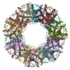
Mass: 40.078 Da / Num. of mol.: 28 / Source method: obtained synthetically / Formula: Ca
#3: Water ChemComp-HOH / water


Mass: 18.015 Da / Num. of mol.: 881 / Source method: isolated from a natural source / Formula: H2O

-
Experimental details

-
Experiment

ExperimentMethod: X-RAY DIFFRACTION / Number of used crystals: 1

-
Sample preparation

CrystalDensity Matthews: 3.48 Å3/Da / Density % sol: 64.63 %
Crystal growTemperature: 298 K / Method: vapor diffusion, sitting drop / pH: 7.4
Details: PEG 6000, CaCl2, MES, pH 7.4, sitting drop, temperature 298.0K, VAPOR DIFFUSION, SITTING DROP

-
Data collection

DiffractionMean temperature: 100 K
Diffraction sourceSource: SYNCHROTRON / Site: SRS / Beamline: PX14.1 / Wavelength: 1.488 Å
DetectorType: ADSC / Detector: CCD / Date: May 6, 2003
RadiationProtocol: SINGLE WAVELENGTH / Scattering type: x-ray
Radiation wavelengthWavelength: 1.488 Å / Relative weight: 1
ReflectionResolution: 2.3→45.27 Å / Num. all: 180419 / Num. obs: 180419 / % possible obs: 89.7 % / Redundancy: 2.1 % / Rmerge(I) obs: 0.08 / Rsym value: 0.08 / Net I/σ(I): 5.435
Reflection shellResolution: 2.3→2.42 Å / Redundancy: 2 % / Rmerge(I) obs: 0.167 / Mean I/σ(I) obs: 3.9 / Num. measured all: 54093 / Num. unique all: 27379 / Rsym value: 0.167 / % possible all: 93.2

-
Phasing

PhasingMethod: molecular replacement
Phasing MR
Highest resolutionLowest resolution
Rotation4 Å20 Å

-
Processing

Software
NameVersionClassificationNB
MOSFLMdata reduction
SCALA3.1.4data scaling
AMoREphasing
CNSrefinement
PDB_EXTRACT3.006data extraction
RefinementMethod to determine structure: MOLECULAR REPLACEMENT
Starting model: Dimer created from A subunit of native octameric Limulus SAP

Resolution: 2.3→45.27 Å / Occupancy max: 1 / Occupancy min: 1 / FOM work R set: 0.853 / Cross valid method: THROUGHOUT / σ(F): 0 / Stereochemistry target values: Engh & Huber / Details: Used maximum likelihood refinement on F
RfactorNum. reflection% reflectionSelection details
Rfree0.223 9112 4.5 %Random
Rwork0.201 ---
all0.219 180238 --
obs0.219 180238 89.4 %-
Solvent computationBsol: 17.258 Å2
Displacement parametersBiso max: 42.04 Å2 / Biso mean: 14.165 Å2 / Biso min: 1 Å2
Baniso -1Baniso -2Baniso -3
1--0.867 Å20 Å20.081 Å2
2--0.583 Å20 Å2
3---0.284 Å2
Refinement stepCycle: LAST / Resolution: 2.3→45.27 Å
ProteinNucleic acidLigandSolventTotal
Num. atoms23352 0 28 881 24261
Refine LS restraints
Refine-IDTypeDev idealDev ideal target
X-RAY DIFFRACTIONc_mcbond_it1.031.5
X-RAY DIFFRACTIONc_scbond_it1.9072
X-RAY DIFFRACTIONc_mcangle_it1.6382
X-RAY DIFFRACTIONc_scangle_it2.6052.5
Refine LS restraints NCS
Ens-IDDom-IDAuth asym-IDRefine-IDRmsTypeWeight
11BX-RAY DIFFRACTION0.02restrain300
11CX-RAY DIFFRACTION0.019restrain300
11DX-RAY DIFFRACTION0.021restrain300
11EX-RAY DIFFRACTION0.018restrain300
11FX-RAY DIFFRACTION0.019restrain300
11GX-RAY DIFFRACTION0.021restrain300
11HX-RAY DIFFRACTION0.02restrain300
11IX-RAY DIFFRACTION0.019restrain300
11JX-RAY DIFFRACTION0.018restrain300
11KX-RAY DIFFRACTION0.02restrain300
11LX-RAY DIFFRACTION0.019restrain300
11MX-RAY DIFFRACTION0.02restrain300
11NX-RAY DIFFRACTION0.022restrain300
LS refinement shellResolution: 2.3→2.38 Å
RfactorNum. reflection
Rfree0.2623 978
Rwork0.2266 -
obs-18715
Xplor file
Refine-IDSerial noParam file
X-RAY DIFFRACTION1CNS_TOPPAR:protein_rep.param
X-RAY DIFFRACTION2CNS_TOPPAR:ion.param
X-RAY DIFFRACTION3CNS_TOPPAR:water_rep.param
X-RAY DIFFRACTION4cis_peptide.param

+
About Yorodumi

-
News

-
Feb 9, 2022. New format data for meta-information of EMDB entries

New format data for meta-information of EMDB entries

  • Version 3 of the EMDB header file is now the official format.
  • The previous official version 1.9 will be removed from the archive.

Related info.:EMDB header

External links:wwPDB to switch to version 3 of the EMDB data model

-
Aug 12, 2020. Covid-19 info

Covid-19 info

URL: https://pdbjlvh1.pdbj.org/emnavi/covid19.php

New page: Covid-19 featured information page in EM Navigator.

Related info.:Covid-19 info / Mar 5, 2020. Novel coronavirus structure data

+
Mar 5, 2020. Novel coronavirus structure data

Novel coronavirus structure data

Related info.:Yorodumi Speices / Aug 12, 2020. Covid-19 info

External links:COVID-19 featured content - PDBj / Molecule of the Month (242):Coronavirus Proteases

+
Jan 31, 2019. EMDB accession codes are about to change! (news from PDBe EMDB page)

EMDB accession codes are about to change! (news from PDBe EMDB page)

  • The allocation of 4 digits for EMDB accession codes will soon come to an end. Whilst these codes will remain in use, new EMDB accession codes will include an additional digit and will expand incrementally as the available range of codes is exhausted. The current 4-digit format prefixed with “EMD-” (i.e. EMD-XXXX) will advance to a 5-digit format (i.e. EMD-XXXXX), and so on. It is currently estimated that the 4-digit codes will be depleted around Spring 2019, at which point the 5-digit format will come into force.
  • The EM Navigator/Yorodumi systems omit the EMD- prefix.

Related info.:Q: What is EMD? / ID/Accession-code notation in Yorodumi/EM Navigator

External links:EMDB Accession Codes are Changing Soon! / Contact to PDBj

+
Jul 12, 2017. Major update of PDB

Major update of PDB

  • wwPDB released updated PDB data conforming to the new PDBx/mmCIF dictionary.
  • This is a major update changing the version number from 4 to 5, and with Remediation, in which all the entries are updated.
  • In this update, many items about electron microscopy experimental information are reorganized (e.g. em_software).
  • Now, EM Navigator and Yorodumi are based on the updated data.

External links:wwPDB Remediation / Enriched Model Files Conforming to OneDep Data Standards Now Available in the PDB FTP Archive

-
Yorodumi

Thousand views of thousand structures

  • Yorodumi is a browser for structure data from EMDB, PDB, SASBDB, etc.
  • This page is also the successor to EM Navigator detail page, and also detail information page/front-end page for Omokage search.
  • The word "yorodu" (or yorozu) is an old Japanese word meaning "ten thousand". "mi" (miru) is to see.

Related info.:EMDB / PDB / SASBDB / Comparison of 3 databanks / Yorodumi Search / Aug 31, 2016. New EM Navigator & Yorodumi / Yorodumi Papers / Jmol/JSmol / Function and homology information / Changes in new EM Navigator and Yorodumi

Read more