[English] 日本語
 Yorodumi
- EMDB-31089: Cryo-EM structure of the octameric state of C-phycocyanin from Th... -
+ Open data
- Basic information
| Entry | Database: EMDB / ID: EMD-31089 | |||||||||||||||
|---|---|---|---|---|---|---|---|---|---|---|---|---|---|---|---|---|
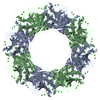
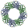
| Title | Cryo-EM structure of the octameric state of C-phycocyanin from Thermoleptolyngbya sp. O-77 | |||||||||||||||
 Map data Map data | ||||||||||||||||
 Sample Sample |
| |||||||||||||||
| Function / homology |  Function and homology information Function and homology information: / phycobilisome / plasma membrane-derived thylakoid membrane / photosynthesis Similarity search - Function | |||||||||||||||
| Biological species |  Leptolyngbya sp. O-77 (bacteria) Leptolyngbya sp. O-77 (bacteria) | |||||||||||||||
| Method | single particle reconstruction / cryo EM / Resolution: 3.71 Å | |||||||||||||||
 Authors Authors | Minato T / Teramoto T / Adachi N / Hung NK / Yamada K / Kawasaki M / Akutsu M / Moriya T / Senda T / Ogo S ...Minato T / Teramoto T / Adachi N / Hung NK / Yamada K / Kawasaki M / Akutsu M / Moriya T / Senda T / Ogo S / Kakuta Y / Yoon KS | |||||||||||||||
| Funding support |  Japan, 4 items Japan, 4 items
| |||||||||||||||
 Citation Citation |  Journal: Commun Biol / Year: 2021 Journal: Commun Biol / Year: 2021Title: Non-conventional octameric structure of C-phycocyanin. Authors: Takuo Minato / Takamasa Teramoto / Naruhiko Adachi / Nguyen Khac Hung / Kaho Yamada / Masato Kawasaki / Masato Akutsu / Toshio Moriya / Toshiya Senda / Seiji Ogo / Yoshimitsu Kakuta / Ki-Seok Yoon /  Abstract: C-phycocyanin (CPC), a blue pigment protein, is an indispensable component of giant phycobilisomes, which are light-harvesting antenna complexes in cyanobacteria that transfer energy efficiently to ...C-phycocyanin (CPC), a blue pigment protein, is an indispensable component of giant phycobilisomes, which are light-harvesting antenna complexes in cyanobacteria that transfer energy efficiently to photosystems I and II. X-ray crystallographic and electron microscopy (EM) analyses have revealed the structure of CPC to be a closed toroidal hexamer by assembling two trimers. In this study, the structural characterization of non-conventional octameric CPC is reported for the first time. Analyses of the crystal and cryogenic EM structures of the native CPC from filamentous thermophilic cyanobacterium Thermoleptolyngbya sp. O-77 unexpectedly illustrated the coexistence of conventional hexamer and novel octamer. In addition, an unusual dimeric state, observed via analytical ultracentrifugation, was postulated to be a key intermediate structure in the assemble of the previously unobserved octamer. These observations provide new insights into the assembly processes of CPCs and the mechanism of energy transfer in the light-harvesting complexes. | |||||||||||||||
| History |
|
- Structure visualization
| Movie |
 Movie viewer Movie viewer |
|---|---|
| Structure viewer | EM map:  SurfView SurfView Molmil Molmil Jmol/JSmol Jmol/JSmol |
| Supplemental images |
- Downloads & links
-EMDB archive
| Map data |  emd_31089.map.gz emd_31089.map.gz | 219.4 MB |  EMDB map data format EMDB map data format | |
|---|---|---|---|---|
| Header (meta data) |  emd-31089-v30.xml emd-31089-v30.xml emd-31089.xml emd-31089.xml | 18.5 KB 18.5 KB | Display Display |  EMDB header EMDB header |
| FSC (resolution estimation) |  emd_31089_fsc.xml emd_31089_fsc.xml | 14.2 KB | Display |  FSC data file FSC data file |
| Images |  emd_31089.png emd_31089.png | 196.7 KB | ||
| Masks |  emd_31089_msk_1.map emd_31089_msk_1.map | 244.1 MB |  Mask map Mask map | |
| Others |  emd_31089_half_map_1.map.gz emd_31089_half_map_1.map.gz emd_31089_half_map_2.map.gz emd_31089_half_map_2.map.gz | 189.5 MB 189.5 MB | ||
| Archive directory |  http://ftp.pdbj.org/pub/emdb/structures/EMD-31089 http://ftp.pdbj.org/pub/emdb/structures/EMD-31089 ftp://ftp.pdbj.org/pub/emdb/structures/EMD-31089 ftp://ftp.pdbj.org/pub/emdb/structures/EMD-31089 | HTTPS FTP |
-Validation report
| Summary document |  emd_31089_validation.pdf.gz emd_31089_validation.pdf.gz | 576.8 KB | Display |  EMDB validaton report EMDB validaton report |
|---|---|---|---|---|
| Full document |  emd_31089_full_validation.pdf.gz emd_31089_full_validation.pdf.gz | 576.3 KB | Display | |
| Data in XML |  emd_31089_validation.xml.gz emd_31089_validation.xml.gz | 21.5 KB | Display | |
| Data in CIF |  emd_31089_validation.cif.gz emd_31089_validation.cif.gz | 28 KB | Display | |
| Arichive directory |  https://ftp.pdbj.org/pub/emdb/validation_reports/EMD-31089 https://ftp.pdbj.org/pub/emdb/validation_reports/EMD-31089 ftp://ftp.pdbj.org/pub/emdb/validation_reports/EMD-31089 ftp://ftp.pdbj.org/pub/emdb/validation_reports/EMD-31089 | HTTPS FTP |
-Related structure data
| Related structure data |  7eh7MC  7efvC  7efwC  7eh8C M: atomic model generated by this map C: citing same article ( |
|---|---|
| Similar structure data | |
| EM raw data |  EMPIAR-11079 (Title: Non-conventional octameric structure of C-phycocyanin EMPIAR-11079 (Title: Non-conventional octameric structure of C-phycocyaninData size: 1.8 TB Data #1: Non-conventional octameric structure of C-phycocyanin [micrographs - multiframe]) |
- Links
Links
| EMDB pages |  EMDB (EBI/PDBe) / EMDB (EBI/PDBe) /  EMDataResource EMDataResource |
|---|
- Map
Map
| File |  Download / File: emd_31089.map.gz / Format: CCP4 / Size: 244.1 MB / Type: IMAGE STORED AS FLOATING POINT NUMBER (4 BYTES) Download / File: emd_31089.map.gz / Format: CCP4 / Size: 244.1 MB / Type: IMAGE STORED AS FLOATING POINT NUMBER (4 BYTES) | ||||||||||||||||||||||||||||||||||||||||||||||||||||||||||||||||||||
|---|---|---|---|---|---|---|---|---|---|---|---|---|---|---|---|---|---|---|---|---|---|---|---|---|---|---|---|---|---|---|---|---|---|---|---|---|---|---|---|---|---|---|---|---|---|---|---|---|---|---|---|---|---|---|---|---|---|---|---|---|---|---|---|---|---|---|---|---|---|
| Projections & slices | Image control
Images are generated by Spider. | ||||||||||||||||||||||||||||||||||||||||||||||||||||||||||||||||||||
| Voxel size | X=Y=Z: 0.88 Å | ||||||||||||||||||||||||||||||||||||||||||||||||||||||||||||||||||||
| Density |
| ||||||||||||||||||||||||||||||||||||||||||||||||||||||||||||||||||||
| Symmetry | Space group: 1 | ||||||||||||||||||||||||||||||||||||||||||||||||||||||||||||||||||||
| Details | EMDB XML:
CCP4 map header:
| ||||||||||||||||||||||||||||||||||||||||||||||||||||||||||||||||||||
-Supplemental data
-Mask #1
| File |  emd_31089_msk_1.map emd_31089_msk_1.map | ||||||||||||
|---|---|---|---|---|---|---|---|---|---|---|---|---|---|
| Projections & Slices |
| ||||||||||||
| Density Histograms |
-Half map: #1
| File | emd_31089_half_map_1.map | ||||||||||||
|---|---|---|---|---|---|---|---|---|---|---|---|---|---|
| Projections & Slices |
| ||||||||||||
| Density Histograms |
-Half map: #2
| File | emd_31089_half_map_2.map | ||||||||||||
|---|---|---|---|---|---|---|---|---|---|---|---|---|---|
| Projections & Slices |
| ||||||||||||
| Density Histograms |
- Sample components
-Entire : CPC octamer
| Entire | Name: CPC octamer |
|---|---|
| Components |
|
-Supramolecule #1: CPC octamer
| Supramolecule | Name: CPC octamer / type: complex / ID: 1 / Parent: 0 / Macromolecule list: all / Details: homo octamer |
|---|---|
| Source (natural) | Organism:  Leptolyngbya sp. O-77 (bacteria) Leptolyngbya sp. O-77 (bacteria) |
| Molecular weight | Theoretical: 320 KDa |
-Macromolecule #1: C-phycocyanin alpha chain
| Macromolecule | Name: C-phycocyanin alpha chain / type: protein_or_peptide / ID: 1 / Enantiomer: LEVO |
|---|---|
| Source (natural) | Organism:  Leptolyngbya sp. O-77 (bacteria) Leptolyngbya sp. O-77 (bacteria) |
| Sequence | String: MKTPITEAIA AADTQGRFLS NTELQAVNGR FERAAASMEA ARALTNNAQQ LIDGAANAVY QKFPYTTQM QGANFASDSR GKSKCARDIG YYLRIITYSL VAGGTGPLDE YLIAGLDEIN R TFDLSPSW YVEALKYIKA NHGLSGQAAN EANTYIDYAI NALS |
-Macromolecule #2: C-phycocyanin beta chain
| Macromolecule | Name: C-phycocyanin beta chain / type: protein_or_peptide / ID: 2 / Details: MEN: N-METHYL ASPARAGINE / Enantiomer: LEVO |
|---|---|
| Source (natural) | Organism:  Leptolyngbya sp. O-77 (bacteria) Leptolyngbya sp. O-77 (bacteria) |
| Sequence | String: MLDAFAKVVS QADTKGEFLS SAQLDALSNV VKDGSKRLDA VNRMTSNAST IVANAARSLF EEQPQLIQP GG(MEN)AYTNRRM AACLRDMEII LRYVTYATLA GDSSVLDDRC LNGLRETYQA L GVPGGSVA AGVAKMKDAA IAIVNDPNGI TKGDCSALVS EIASYFDRAA AAVA |
-Experimental details
-Structure determination
| Method | cryo EM |
|---|---|
 Processing Processing | single particle reconstruction |
| Aggregation state | particle |
- Sample preparation
| Concentration | 6.7 mg/mL |
|---|---|
| Buffer | pH: 7 |
| Grid | Model: Quantifoil R1.2/1.3 / Material: COPPER / Mesh: 300 / Support film - Material: CARBON / Support film - topology: HOLEY / Pretreatment - Type: GLOW DISCHARGE / Pretreatment - Atmosphere: AIR / Details: The grid was washed by acetone prior to use. |
| Vitrification | Cryogen name: ETHANE / Chamber humidity: 100 % / Chamber temperature: 291 K / Instrument: FEI VITROBOT MARK IV / Details: Blotting time was 5 seconds (blot force 15). |
| Details | This sample was mono-disperse. |
- Electron microscopy
| Microscope | TFS TALOS |
|---|---|
| Image recording | Film or detector model: FEI FALCON III (4k x 4k) / Detector mode: COUNTING / Number grids imaged: 2 / Number real images: 2036 / Average exposure time: 49.2 sec. / Average electron dose: 50.0 e/Å2 |
| Electron beam | Acceleration voltage: 200 kV / Electron source:  FIELD EMISSION GUN FIELD EMISSION GUN |
| Electron optics | C2 aperture diameter: 50.0 µm / Illumination mode: FLOOD BEAM / Imaging mode: BRIGHT FIELD / Cs: 2.7 mm / Nominal defocus max: 2.5 µm / Nominal defocus min: 1.0 µm / Nominal magnification: 120000 |
| Sample stage | Cooling holder cryogen: NITROGEN |
 Movie
Movie Controller


 Z (Sec.)
 Y (Row.)
 X (Col.)


