 Movie
Movie Controller


+ Open data

- Basic information
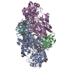
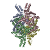
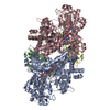
| Entry | Database: PDB / ID: 7ebe | ||||||
|---|---|---|---|---|---|---|---|
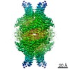
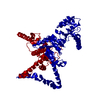
| Title | Crystal structure of Isocitrate lyase-1 from Candida albicans | ||||||
 Components Components | Isocitrate lyase | ||||||
 Keywords Keywords | LYASE / Isocitrate lyase / glyoxylate cycle / ubiquitination | ||||||
| Function / homology |  Function and homology information Function and homology informationisocitrate lyase activity / methylisocitrate lyase activity / glyoxylate cycle / tricarboxylic acid cycle / peroxisome / metal ion binding Similarity search - Function | ||||||
| Biological species |  Candida albicans (yeast) Candida albicans (yeast) | ||||||
| Method |  X-RAY DIFFRACTION / SYNCHROTRON / MOLECULAR REPLACEMENT / Resolution: 2.69 Å X-RAY DIFFRACTION / SYNCHROTRON / MOLECULAR REPLACEMENT / Resolution: 2.69 Å | ||||||
 Authors Authors | Hiragi, K. / Nishio, K. / Moriyama, S. / Hamaguchi, T. / Mizoguchi, A. / Yonekura, K. / Tani, K. / Mizushima, T. | ||||||
 Citation Citation | Journal: J Struct Biol / Year: 2021 Title: Structural insights into the targeting specificity of ubiquitin ligase for S. cerevisiae isocitrate lyase but not C. albicans isocitrate lyase. Authors: Keito Hiragi / Kazuya Nishio / Shu Moriyama / Tasuku Hamaguchi / Akira Mizoguchi / Koji Yonekura / Kazutoshi Tani / Tsunehiro Mizushima /  Abstract: In Saccharomyces cerevisiae, the glyoxylate cycle is controlled through the posttranslational regulation of its component enzymes, such as isocitrate lyase (ICL), which catalyzes the first unique ...In Saccharomyces cerevisiae, the glyoxylate cycle is controlled through the posttranslational regulation of its component enzymes, such as isocitrate lyase (ICL), which catalyzes the first unique step of the cycle. The ICL of S.cerevisiae (ScIcl1) is tagged for proteasomal degradation through ubiquitination by a multisubunit ubiquitin ligase (the glucose-induced degradation-deficient (GID) complex), whereas that of the pathogenic yeast Candida albicans (CaIcl1) escapes this process. However, the reason for the ubiquitin targeting specificity of the GID complex for ScIcl1 and not for CaIcl1 is unclear. To gain some insight into this, in this study, the crystal structures of apo ScIcl1 and CaIcl1 in complex with formate and the cryogenic electron microscopy structure of apo CaIcl1 were determined at a resolution of 2.3, 2.7, and 2.6 Å, respectively. A comparison of the various structures suggests that the orientation of N-terminal helix α1 in S.cerevisiae is likely key to repositioning of ubiquitination sites and contributes to the distinction found in C. albicans ubiquitin evasion mechanism. This finding gives us a better understanding of the molecular mechanism of ubiquitin-dependent ScIcl1 degradation and could serve as a theoretical basis for the research and development of anti-C. albicans drugs based on the concept of CaIcl1 ubiquitination. | ||||||
| History |
|
- Structure visualization
| Structure viewer | Molecule: MolmilJmol/JSmol |
|---|
- Downloads & links
-Download
| PDBx/mmCIF format | 7ebe.cif.gz | 827 KB | Display | PDBx/mmCIF format |
|---|---|---|---|---|
| PDB format | pdb7ebe.ent.gz | 685.8 KB | Display | PDB format |
| PDBx/mmJSON format | 7ebe.json.gz | Tree view | PDBx/mmJSON format | |
| Others |  Other downloads Other downloads |
-Validation report
| Arichive directory | https://data.pdbj.org/pub/pdb/validation_reports/eb/7ebeftp://data.pdbj.org/pub/pdb/validation_reports/eb/7ebe | HTTPS FTP |
|---|
-Related structure data
| Related structure data | 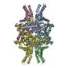 7ebcSC 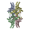 7ebfC S: Starting model for refinement C: citing same article ( |
|---|---|
| Similar structure data |


-Links
 PDBj
PDBj
- Assembly
| Deposited unit | 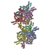
| |||||||||||||||||||||||||||||||||||||||||||||||||||||||||||||||||||||||||||||||||||||||||||||||||||||||||||||||||||||||||||||||||||||||||||||||||||||||||||||||||||||||||||||||||||||||||||||||||||||||||||||||||||||||||||||||||||||||||||||||||||||||||||||||||||||||||||||||||||||||||||||||||||||||||||||||||||||
|---|---|---|---|---|---|---|---|---|---|---|---|---|---|---|---|---|---|---|---|---|---|---|---|---|---|---|---|---|---|---|---|---|---|---|---|---|---|---|---|---|---|---|---|---|---|---|---|---|---|---|---|---|---|---|---|---|---|---|---|---|---|---|---|---|---|---|---|---|---|---|---|---|---|---|---|---|---|---|---|---|---|---|---|---|---|---|---|---|---|---|---|---|---|---|---|---|---|---|---|---|---|---|---|---|---|---|---|---|---|---|---|---|---|---|---|---|---|---|---|---|---|---|---|---|---|---|---|---|---|---|---|---|---|---|---|---|---|---|---|---|---|---|---|---|---|---|---|---|---|---|---|---|---|---|---|---|---|---|---|---|---|---|---|---|---|---|---|---|---|---|---|---|---|---|---|---|---|---|---|---|---|---|---|---|---|---|---|---|---|---|---|---|---|---|---|---|---|---|---|---|---|---|---|---|---|---|---|---|---|---|---|---|---|---|---|---|---|---|---|---|---|---|---|---|---|---|---|---|---|---|---|---|---|---|---|---|---|---|---|---|---|---|---|---|---|---|---|---|---|---|---|---|---|---|---|---|---|---|---|---|---|---|---|---|---|---|---|---|---|---|---|---|---|---|---|---|---|---|---|---|---|---|---|---|---|---|---|---|---|---|---|---|---|---|---|---|---|---|---|---|---|---|---|---|---|---|---|---|---|---|
| 1 | 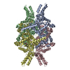
| |||||||||||||||||||||||||||||||||||||||||||||||||||||||||||||||||||||||||||||||||||||||||||||||||||||||||||||||||||||||||||||||||||||||||||||||||||||||||||||||||||||||||||||||||||||||||||||||||||||||||||||||||||||||||||||||||||||||||||||||||||||||||||||||||||||||||||||||||||||||||||||||||||||||||||||||||||||
| 2 | 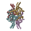
| |||||||||||||||||||||||||||||||||||||||||||||||||||||||||||||||||||||||||||||||||||||||||||||||||||||||||||||||||||||||||||||||||||||||||||||||||||||||||||||||||||||||||||||||||||||||||||||||||||||||||||||||||||||||||||||||||||||||||||||||||||||||||||||||||||||||||||||||||||||||||||||||||||||||||||||||||||||
| Unit cell |
| |||||||||||||||||||||||||||||||||||||||||||||||||||||||||||||||||||||||||||||||||||||||||||||||||||||||||||||||||||||||||||||||||||||||||||||||||||||||||||||||||||||||||||||||||||||||||||||||||||||||||||||||||||||||||||||||||||||||||||||||||||||||||||||||||||||||||||||||||||||||||||||||||||||||||||||||||||||
| Noncrystallographic symmetry (NCS) | NCS domain:
NCS domain segments: Component-ID: _ / Refine code: _
|