 Movie
Movie Controller

[English] 日本語
 Yorodumi
- PDB-1dqu: CRYSTAL STRUCTURE OF THE ISOCITRATE LYASE FROM ASPERGILLUS NIDULANS -
+ Open data

- Basic information
| Entry | Database: PDB / ID: 1dqu | ||||||
|---|---|---|---|---|---|---|---|
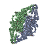
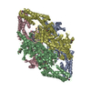
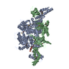
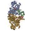
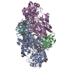
| Title | CRYSTAL STRUCTURE OF THE ISOCITRATE LYASE FROM ASPERGILLUS NIDULANS | ||||||
 Components Components | ISOCITRATE LYASE | ||||||
 Keywords Keywords | LYASE / BETA BARREL | ||||||
| Function / homology |  Function and homology information Function and homology informationglyoxysome / acetate catabolic process / methylisocitrate lyase / isocitrate lyase / isocitrate lyase activity / methylisocitrate lyase activity / carbon utilization / fatty acid catabolic process / glyoxylate cycle / peroxisomal matrix ...glyoxysome / acetate catabolic process / methylisocitrate lyase / isocitrate lyase / isocitrate lyase activity / methylisocitrate lyase activity / carbon utilization / fatty acid catabolic process / glyoxylate cycle / peroxisomal matrix / tricarboxylic acid cycle / peroxisome / metal ion binding Similarity search - Function | ||||||
| Biological species |  | ||||||
| Method |  X-RAY DIFFRACTION / SYNCHROTRON / Resolution: 2.8 Å X-RAY DIFFRACTION / SYNCHROTRON / Resolution: 2.8 Å | ||||||
 Authors Authors | Britton, K.L. / Langridge, S.J. / Baker, P.J. / Weeradechapon, K. / Sedelnikova, S.E. / De Lucas, J.R. / Rice, D.W. / Turner, G. | ||||||
 Citation Citation | Journal: Structure Fold.Des. / Year: 2000 Title: The crystal structure and active site location of isocitrate lyase from the fungus Aspergillus nidulans. Authors: Britton, K. / Langridge, S. / Baker, P.J. / Weeradechapon, K. / Sedelnikova, S.E. / De Lucas, J.R. / Rice, D.W. / Turner, G. #1: Journal: Acta Crystallogr.,Sect.D / Year: 1997Title: Isocitrate Lyase from Aspergillus Nidulans: Crystallization and X-ray Analysis of a Glyoxylate Cycle Enzyme. Authors: Langridge, S.J. / Baker, P.J. / De Lucas, J.R. / Sedelnikova, S.E. / Turner, G. / Rice, D.W. | ||||||
| History |
|
- Structure visualization
| Structure viewer | Molecule: MolmilJmol/JSmol |
|---|
- Downloads & links
-Download
| PDBx/mmCIF format | 1dqu.cif.gz | 97.9 KB | Display | PDBx/mmCIF format |
|---|---|---|---|---|
| PDB format | pdb1dqu.ent.gz | 74.5 KB | Display | PDB format |
| PDBx/mmJSON format | 1dqu.json.gz | Tree view | PDBx/mmJSON format | |
| Others |  Other downloads Other downloads |
-Validation report
| Arichive directory | https://data.pdbj.org/pub/pdb/validation_reports/dq/1dquftp://data.pdbj.org/pub/pdb/validation_reports/dq/1dqu | HTTPS FTP |
|---|
-Related structure data
| Similar structure data |
|---|


-Links
 PDBj
PDBj
- Assembly
| Deposited unit | 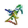
| ||||||||
|---|---|---|---|---|---|---|---|---|---|
| 1 | 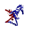
| ||||||||
| Unit cell |
| ||||||||
| Details | The biological assembly is a tetramer constructed from chain A generated by 222 symmetry |
-Components
| #1: Protein | Mass: 60398.336 Da / Num. of mol.: 1 Source method: isolated from a genetically manipulated source Source: (gene. exp.) |
|---|
-Experimental details
-Experiment
| Experiment | Method: X-RAY DIFFRACTION / Number of used crystals: 1 |
|---|
- Sample preparation
| Crystal | Density Matthews: 2.67 Å3/Da / Density % sol: 53.95 % | ||||||||||||||||||||
|---|---|---|---|---|---|---|---|---|---|---|---|---|---|---|---|---|---|---|---|---|---|
| Crystal grow | Temperature: 290 K / Method: vapor diffusion, hanging drop / pH: 7.5 Details: PEG 2000, sodium HEPES, pH 7.5, VAPOR DIFFUSION, HANGING DROP, temperature 290K | ||||||||||||||||||||
| Crystal grow | *PLUS Details: Langridge, S.J., (1997) Acta Crystallogr., Sect.D, 53, 488. | ||||||||||||||||||||
| Components of the solutions | *PLUS
|
-Data collection
| Diffraction | Mean temperature: 298 K |
|---|---|
| Diffraction source | Source: SYNCHROTRON / Site: SRS  / Beamline: PX9.6 / Wavelength: 0.87 / Beamline: PX9.6 / Wavelength: 0.87 |
| Detector | Type: MARRESEARCH / Detector: IMAGE PLATE / Date: Feb 27, 1996 |
| Radiation | Protocol: SINGLE WAVELENGTH / Monochromatic (M) / Laue (L): M / Scattering type: x-ray |
| Radiation wavelength | Wavelength: 0.87 Å / Relative weight: 1 |
| Reflection | Resolution: 2.5→20 Å / Num. all: 21252 / Num. obs: 21252 / % possible obs: 91.5 % / Redundancy: 2.9 % / Rmerge(I) obs: 0.05 / Net I/σ(I): 13.2 |
| Reflection shell | Resolution: 2.5→2.56 Å / Redundancy: 2.2 % / Rmerge(I) obs: 0.439 / Num. unique all: 1198 / % possible all: 71.9 |
| Reflection | *PLUS Num. measured all: 62390 |
- Processing
| Software |
| |||||||||||||||||||||||||
|---|---|---|---|---|---|---|---|---|---|---|---|---|---|---|---|---|---|---|---|---|---|---|---|---|---|---|
| Refinement | Resolution: 2.8→10 Å / Stereochemistry target values: Engh & Huber
| |||||||||||||||||||||||||
| Refinement step | Cycle: LAST / Resolution: 2.8→10 Å
| |||||||||||||||||||||||||
| Refine LS restraints |
| |||||||||||||||||||||||||
| Software | *PLUS Name: TNT / Classification: refinement | |||||||||||||||||||||||||
| Refinement | *PLUS Highest resolution: 2.8 Å / Rfactor all: 0.276 | |||||||||||||||||||||||||
| Solvent computation | *PLUS | |||||||||||||||||||||||||
| Displacement parameters | *PLUS | |||||||||||||||||||||||||
| Refine LS restraints | *PLUS
|
