 Movie
Movie Controller

[English] 日本語
 Yorodumi
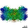
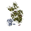
- PDB-7dsx: Structure of a human NHE1-CHP1 complex under pH 7.5, bound by car... -
+ Open data

- Basic information
| Entry | Database: PDB / ID: 7dsx | ||||||
|---|---|---|---|---|---|---|---|
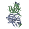
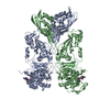
| Title | Structure of a human NHE1-CHP1 complex under pH 7.5, bound by cariporide | ||||||
 Components Components |
| ||||||
 Keywords Keywords | MEMBRANE PROTEIN / Transporter | ||||||
| Function / homology |  Function and homology information Function and homology informationnegative regulation of phosphatase activity / positive regulation of sodium:proton antiporter activity / Sodium/Proton exchangers / regulation of the force of heart contraction by cardiac conduction / cation-transporting ATPase complex / membrane docking / negative regulation of protein autophosphorylation / Hyaluronan degradation / positive regulation of protein transport / regulation of cardiac muscle cell membrane potential ...negative regulation of phosphatase activity / positive regulation of sodium:proton antiporter activity / Sodium/Proton exchangers / regulation of the force of heart contraction by cardiac conduction / cation-transporting ATPase complex / membrane docking / negative regulation of protein autophosphorylation / Hyaluronan degradation / positive regulation of protein transport / regulation of cardiac muscle cell membrane potential / transporter complex / positive regulation of phospholipid biosynthetic process / cellular response to electrical stimulus / potassium:proton antiporter activity / sodium:proton antiporter activity / positive regulation of action potential / hyaluronan catabolic process / positive regulation of glycoprotein biosynthetic process / maintenance of cell polarity / regulation of pH / positive regulation of calcineurin-NFAT signaling cascade / sodium ion export across plasma membrane / membrane organization / protein phosphatase 2B binding / microtubule bundle formation / intracellular sodium ion homeostasis / regulation of stress fiber assembly / cardiac muscle cell differentiation / positive regulation of mitochondrial membrane permeability / sodium ion import across plasma membrane / : / negative regulation of calcineurin-NFAT signaling cascade / regulation of cardiac muscle contraction by calcium ion signaling / regulation of focal adhesion assembly / response to acidic pH / cardiac muscle cell contraction / negative regulation of protein import into nucleus / small GTPase-mediated signal transduction / cellular response to cold / positive regulation of cardiac muscle hypertrophy / positive regulation of the force of heart contraction / negative regulation of protein phosphorylation / endoplasmic reticulum-Golgi intermediate compartment / cellular response to antibiotic / protein kinase inhibitor activity / protein complex oligomerization / intercalated disc / potassium channel regulator activity / positive regulation of protein targeting to membrane / negative regulation of protein kinase activity / transport vesicle / cellular response to acidic pH / response to muscle stretch / cytoplasmic microtubule organization / phosphatidylinositol-4,5-bisphosphate binding / monoatomic ion transport / protein export from nucleus / cellular response to epinephrine stimulus / negative regulation of protein ubiquitination / T-tubule / potassium ion transmembrane transport / proton transmembrane transport / regulation of intracellular pH / stem cell differentiation / potassium ion transport / cellular response to mechanical stimulus / phospholipid binding / kinase binding / cellular response to insulin stimulus / calcium-dependent protein binding / cell migration / lamellipodium / microtubule cytoskeleton / positive regulation of cell growth / cellular response to hypoxia / microtubule binding / molecular adaptor activity / basolateral plasma membrane / membrane fusion / protein-macromolecule adaptor activity / calmodulin binding / apical plasma membrane / protein stabilization / positive regulation of apoptotic process / membrane raft / Golgi membrane / focal adhesion / calcium ion binding / negative regulation of apoptotic process / perinuclear region of cytoplasm / cell surface / endoplasmic reticulum / positive regulation of transcription by RNA polymerase II / mitochondrion / extracellular exosome / nucleoplasm / membrane / identical protein binding / nucleus / plasma membrane Similarity search - Function | ||||||
| Biological species |  Homo sapiens (human) Homo sapiens (human) | ||||||
| Method | ELECTRON MICROSCOPY / single particle reconstruction / cryo EM / Resolution: 3.5 Å | ||||||
 Authors Authors | Dong, Y. / Gao, Y. / Li, B. / Zhang, X.C. / Zhao, Y. | ||||||
 Citation Citation | Journal: Nat Commun / Year: 2021 Title: Structure and mechanism of the human NHE1-CHP1 complex. Authors: Yanli Dong / Yiwei Gao / Alina Ilie / DuSik Kim / Annie Boucher / Bin Li / Xuejun C Zhang / John Orlowski / Yan Zhao /   Abstract: Sodium/proton exchanger 1 (NHE1) is an electroneutral secondary active transporter present on the plasma membrane of most mammalian cells and plays critical roles in regulating intracellular pH and ...Sodium/proton exchanger 1 (NHE1) is an electroneutral secondary active transporter present on the plasma membrane of most mammalian cells and plays critical roles in regulating intracellular pH and volume homeostasis. Calcineurin B-homologous protein 1 (CHP1) is an obligate binding partner that promotes NHE1 biosynthetic maturation, cell surface expression and pH-sensitivity. Dysfunctions of either protein are associated with neurological disorders. Here, we elucidate structures of the human NHE1-CHP1 complex in both inward- and inhibitor (cariporide)-bound outward-facing conformations. We find that NHE1 assembles as a symmetrical homodimer, with each subunit undergoing an elevator-like conformational change during cation exchange. The cryo-EM map reveals the binding site for the NHE1 inhibitor cariporide, illustrating how inhibitors block transport activity. The CHP1 molecule differentially associates with these two conformational states of each NHE1 monomer, and this association difference probably underlies the regulation of NHE1 pH-sensitivity by CHP1. | ||||||
| History |
|
- Structure visualization
| Movie |
Movie viewer |
|---|---|
| Structure viewer | Molecule: MolmilJmol/JSmol |
- Downloads & links
-Download
| PDBx/mmCIF format | 7dsx.cif.gz | 252.1 KB | Display | PDBx/mmCIF format |
|---|---|---|---|---|
| PDB format | pdb7dsx.ent.gz | 200.9 KB | Display | PDB format |
| PDBx/mmJSON format | 7dsx.json.gz | Tree view | PDBx/mmJSON format | |
| Others |  Other downloads Other downloads |
-Validation report
| Arichive directory | https://data.pdbj.org/pub/pdb/validation_reports/ds/7dsxftp://data.pdbj.org/pub/pdb/validation_reports/ds/7dsx | HTTPS FTP |
|---|
-Related structure data
| Related structure data | 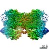 30849MC 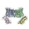 7dsvC 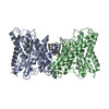 7dswC M: map data used to model this data C: citing same article ( |
|---|---|
| Similar structure data |


-Links
 PDBj
PDBj- Assembly
| Deposited unit | 
|
|---|---|
| 1 |
|
-Components
| #1: Protein | Mass: 21369.830 Da / Num. of mol.: 2 / Mutation: I171A Source method: isolated from a genetically manipulated source Source: (gene. exp.) Homo sapiens (human) / Gene: CHP1, CHP / Cell line (production host): HEK293 / Production host: Homo sapiens (human) / References: UniProt: Q99653#2: Protein | Mass: 57100.363 Da / Num. of mol.: 2 Source method: isolated from a genetically manipulated source Source: (gene. exp.) Homo sapiens (human) / Gene: SLC9A1, APNH1, NHE1 / Cell line (production host): HEK293 / Production host: Homo sapiens (human) / References: UniProt: P19634#3: Chemical | ChemComp-PGT / (   Mass: 751.023 Da / Num. of mol.: 6 / Source method: obtained synthetically / Formula: C40H79O10P / Comment: phospholipid*YM Mass: 751.023 Da / Num. of mol.: 6 / Source method: obtained synthetically / Formula: C40H79O10P / Comment: phospholipid*YM#4: Chemical |   Mass: 283.347 Da / Num. of mol.: 2 / Source method: obtained synthetically / Formula: C12H17N3O3S / Feature type: SUBJECT OF INVESTIGATION Mass: 283.347 Da / Num. of mol.: 2 / Source method: obtained synthetically / Formula: C12H17N3O3S / Feature type: SUBJECT OF INVESTIGATIONHas ligand of interest | Y | |
|---|
-Experimental details
-Experiment
| Experiment | Method: ELECTRON MICROSCOPY |
|---|---|
| EM experiment | Aggregation state: PARTICLE / 3D reconstruction method: single particle reconstruction |
- Sample preparation
| Component |
| ||||||||||||||||||||||||
|---|---|---|---|---|---|---|---|---|---|---|---|---|---|---|---|---|---|---|---|---|---|---|---|---|---|
| Molecular weight |
| ||||||||||||||||||||||||
| Source (natural) |
| ||||||||||||||||||||||||
| Source (recombinant) |
| ||||||||||||||||||||||||
| Buffer solution | pH: 7.5 | ||||||||||||||||||||||||
| Specimen | Conc.: 6 mg/ml / Embedding applied: NO / Shadowing applied: NO / Staining applied: NO / Vitrification applied: YES Details: The NHE1-CHP1 complex was reconstituted into lipid nanodiscs. | ||||||||||||||||||||||||
| Specimen support | Grid material: COPPER / Grid mesh size: 200 divisions/in. / Grid type: Quantifoil | ||||||||||||||||||||||||
| Vitrification | Instrument: FEI VITROBOT MARK III / Cryogen name: ETHANE / Humidity: 100 % / Chamber temperature: 277 K |
- Electron microscopy imaging
| Experimental equipment |  Model: Titan Krios / Image courtesy: FEI Company |
|---|---|
| Microscopy | Model: FEI TITAN KRIOS |
| Electron gun | Electron source:  FIELD EMISSION GUN / Accelerating voltage: 300 kV / Illumination mode: FLOOD BEAM FIELD EMISSION GUN / Accelerating voltage: 300 kV / Illumination mode: FLOOD BEAM |
| Electron lens | Mode: BRIGHT FIELD / Nominal magnification: 13000 X / Nominal defocus max: 2200 nm / Nominal defocus min: 1200 nm / Cs: 2.7 mm / Alignment procedure: COMA FREE |
| Specimen holder | Cryogen: NITROGEN / Specimen holder model: FEI TITAN KRIOS AUTOGRID HOLDER |
| Image recording | Electron dose: 60 e/Å2 / Detector mode: SUPER-RESOLUTION / Film or detector model: GATAN K2 SUMMIT (4k x 4k) / Num. of grids imaged: 1 / Num. of real images: 3855 |
| EM imaging optics | Energyfilter name: GIF Bioquantum / Energyfilter slit width: 20 eV |
| Image scans | Width: 3838 / Height: 3710 / Movie frames/image: 32 / Used frames/image: 1-32 |
- Processing
| EM software |
| ||||||||||||||||||||||||||||||||||||||||||||
|---|---|---|---|---|---|---|---|---|---|---|---|---|---|---|---|---|---|---|---|---|---|---|---|---|---|---|---|---|---|---|---|---|---|---|---|---|---|---|---|---|---|---|---|---|---|
| CTF correction | Type: PHASE FLIPPING AND AMPLITUDE CORRECTION | ||||||||||||||||||||||||||||||||||||||||||||
| 3D reconstruction | Resolution: 3.5 Å / Resolution method: FSC 0.143 CUT-OFF / Num. of particles: 61460 / Symmetry type: POINT | ||||||||||||||||||||||||||||||||||||||||||||
| Atomic model building | Protocol: AB INITIO MODEL |
