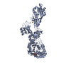
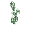
登録情報 データベース : PDB / ID : 6h5oタイトル Crystal structure of PBP2a from MRSA in complex with piperacillin at active site. Penicillin binding protein 2 prime キーワード / 機能・相同性 分子機能 ドメイン・相同性 構成要素
/ / / / / / / / / / / / / / / / / / / / / / / / / / / / / / / / / / / / / / / / 生物種 Staphylococcus aureus (黄色ブドウ球菌)手法 / / / 解像度 : 2.82 Å データ登録者 Batuecas, M.T. / Martinez-Caballero, S. / Hermoso, J.A. 資金援助 組織 認可番号 国 National Institutes of Health/National Institute Of Allergy and Infectious Diseases (NIH/NIAID) 1R01AI116548
ジャーナル : Antimicrob.Agents Chemother. / 年 : 2019タイトル : The Quinazolinone Allosteric Inhibitor of PBP 2a Synergizes with Piperacillin and Tazobactam against Methicillin-Resistant Staphylococcus aureus.著者 : Janardhanan, J. / Bouley, R. / Martinez-Caballero, S. / Peng, Z. / Batuecas-Mordillo, M. / Meisel, J.E. / Ding, D. / Schroeder, V.A. / Wolter, W.R. / Mahasenan, K.V. / Hermoso, J.A. / Mobashery, S. / Chang, M. 履歴 登録 2018年7月25日 登録サイト / 処理サイト 改定 1.0 2019年8月14日 Provider / タイプ 改定 1.1 2020年9月9日 Group / Derived calculationsカテゴリ citation / citation_author ... citation / citation_author / struct_conn / struct_conn_type Item _citation.country / _citation.journal_abbrev ... _citation.country / _citation.journal_abbrev / _citation.journal_id_ASTM / _citation.journal_id_CSD / _citation.journal_id_ISSN / _citation.journal_volume / _citation.pdbx_database_id_DOI / _citation.pdbx_database_id_PubMed / _citation.title / _citation.year / _struct_conn.conn_type_id / _struct_conn.id / _struct_conn.pdbx_dist_value / _struct_conn.pdbx_leaving_atom_flag / _struct_conn.ptnr1_auth_asym_id / _struct_conn.ptnr1_auth_comp_id / _struct_conn.ptnr1_auth_seq_id / _struct_conn.ptnr1_label_asym_id / _struct_conn.ptnr1_label_atom_id / _struct_conn.ptnr1_label_comp_id / _struct_conn.ptnr1_label_seq_id / _struct_conn.ptnr2_auth_asym_id / _struct_conn.ptnr2_auth_comp_id / _struct_conn.ptnr2_auth_seq_id / _struct_conn.ptnr2_label_asym_id / _struct_conn.ptnr2_label_atom_id / _struct_conn.ptnr2_label_comp_id / _struct_conn.ptnr2_label_seq_id / _struct_conn_type.id 改定 1.2 2022年3月30日 Group / Database references / カテゴリ / pdbx_audit_supportItem / _database_2.pdbx_database_accession / _pdbx_audit_support.funding_organization改定 1.3 2024年1月17日 Group / Refinement descriptionカテゴリ chem_comp_atom / chem_comp_bond ... chem_comp_atom / chem_comp_bond / pdbx_initial_refinement_model / struct_ncs_dom_lim Item _struct_ncs_dom_lim.beg_auth_comp_id / _struct_ncs_dom_lim.beg_label_asym_id ... _struct_ncs_dom_lim.beg_auth_comp_id / _struct_ncs_dom_lim.beg_label_asym_id / _struct_ncs_dom_lim.beg_label_comp_id / _struct_ncs_dom_lim.beg_label_seq_id / _struct_ncs_dom_lim.end_auth_comp_id / _struct_ncs_dom_lim.end_label_asym_id / _struct_ncs_dom_lim.end_label_comp_id / _struct_ncs_dom_lim.end_label_seq_id 改定 1.4 2024年10月16日 Group カテゴリ / pdbx_modification_feature
すべて表示 表示を減らす
 ムービー
ムービー コントローラー


 データを開く
 基本情報
基本情報 要素
要素 キーワード
キーワード 機能・相同性情報
 Staphylococcus aureus (黄色ブドウ球菌)
 X線回折 /
  データ登録者
 米国, 1件
  引用
引用 構造の表示
構造の表示 ダウンロードとリンク
 その他のダウンロード


 PDBj
PDBj

 集合体
集合体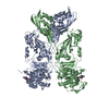
 分子量: 519.571 Da / 分子数: 2 / 由来タイプ: 組換発現 / 式: C23H29N5O7S
 分子量: 112.411 Da / 分子数: 9 / 由来タイプ: 合成 / 式: Cd
 分子量: 18.015 Da / 分子数: 112 / 由来タイプ: 天然 / 式: H2O
 試料調製
試料調製 / ビームライン: XALOC / 波長: 0.97926 Å
 解析
解析