 Movie
Movie Controller

[English] 日本語
 Yorodumi
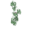
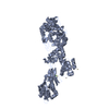
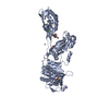
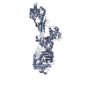
- PDB-6q9n: Crystal structure of PBP2a from MRSA in complex with piperacillin... -
+ Open data

- Basic information
| Entry | Database: PDB / ID: 6q9n | ||||||
|---|---|---|---|---|---|---|---|
| Title | Crystal structure of PBP2a from MRSA in complex with piperacillin and quinazolinone | ||||||
 Components Components | Penicillin binding protein 2 prime | ||||||
 Keywords Keywords | HYDROLASE / Penicillin Binding Protein | ||||||
| Function / homology |  Function and homology information Function and homology informationpeptidoglycan L,D-transpeptidase activity / penicillin binding / cell wall organization / response to antibiotic / plasma membrane Similarity search - Function | ||||||
| Biological species |   Staphylococcus aureus (bacteria) Staphylococcus aureus (bacteria) | ||||||
| Method |  X-RAY DIFFRACTION / SYNCHROTRON / MOLECULAR REPLACEMENT / Resolution: 2.5 Å X-RAY DIFFRACTION / SYNCHROTRON / MOLECULAR REPLACEMENT / Resolution: 2.5 Å | ||||||
 Authors Authors | Martinez-Caballero, S. / Batuecas, M.T. / Hermoso, J.A. | ||||||
| Funding support |  United States, 1items United States, 1items
| ||||||
 Citation Citation | Journal: Antimicrob.Agents Chemother. / Year: 2019 Title: The Quinazolinone Allosteric Inhibitor of PBP 2a Synergizes with Piperacillin and Tazobactam against Methicillin-Resistant Staphylococcus aureus. Authors: Janardhanan, J. / Bouley, R. / Martinez-Caballero, S. / Peng, Z. / Batuecas-Mordillo, M. / Meisel, J.E. / Ding, D. / Schroeder, V.A. / Wolter, W.R. / Mahasenan, K.V. / Hermoso, J.A. / Mobashery, S. / Chang, M. | ||||||
| History |
|
- Structure visualization
| Structure viewer | Molecule: MolmilJmol/JSmol |
|---|
- Downloads & links
-Download
| PDBx/mmCIF format | 6q9n.cif.gz | 270.6 KB | Display | PDBx/mmCIF format |
|---|---|---|---|---|
| PDB format | pdb6q9n.ent.gz | 215.5 KB | Display | PDB format |
| PDBx/mmJSON format | 6q9n.json.gz | Tree view | PDBx/mmJSON format | |
| Others |  Other downloads Other downloads |
-Validation report
| Arichive directory | https://data.pdbj.org/pub/pdb/validation_reports/q9/6q9nftp://data.pdbj.org/pub/pdb/validation_reports/q9/6q9n | HTTPS FTP |
|---|
-Related structure data
| Related structure data | 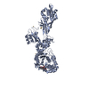 6h5oC  3zg0S S: Starting model for refinement C: citing same article ( |
|---|---|
| Similar structure data |


-Links
 PDBj
PDBj
- Assembly
| Deposited unit | 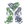
| ||||||||
|---|---|---|---|---|---|---|---|---|---|
| 1 | 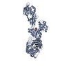
| ||||||||
| 2 | 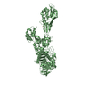
| ||||||||
| Unit cell |
|
-Components
-Protein / Sugars , 2 types, 3 molecules AB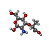
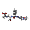
| #1: Protein | Mass: 73286.656 Da / Num. of mol.: 2 Source method: isolated from a genetically manipulated source Details: In chain A, the Ser is attached covalently to piperacillin (PA4 residue 403) Source: (gene. exp.) Staphylococcus aureus (strain Mu50 / ATCC 700699) (bacteria)Strain: Mu50 / ATCC 700699 / Gene: mecA, SAV0041 / Production host: #6: Sugar | ChemComp-MUR / |  Type: D-saccharide, beta linking / Mass: 251.234 Da / Num. of mol.: 1 Type: D-saccharide, beta linking / Mass: 251.234 Da / Num. of mol.: 1Source method: isolated from a genetically manipulated source Formula: C9H17NO7 |
|---|
-Non-polymers , 5 types, 99 molecules 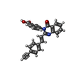


| #2: Chemical | ChemComp-QLN /  Mass: 392.406 Da / Num. of mol.: 1 / Source method: obtained synthetically / Formula: C25H16N2O3 Mass: 392.406 Da / Num. of mol.: 1 / Source method: obtained synthetically / Formula: C25H16N2O3 | ||||
|---|---|---|---|---|---|
| #3: Chemical | ChemComp-JPP /  Mass: 519.571 Da / Num. of mol.: 1 / Source method: obtained synthetically / Formula: C23H29N5O7S Mass: 519.571 Da / Num. of mol.: 1 / Source method: obtained synthetically / Formula: C23H29N5O7S | ||||
| #4: Chemical | ChemComp-CL /  Mass: 35.453 Da / Num. of mol.: 4 / Source method: obtained synthetically / Formula: Cl Mass: 35.453 Da / Num. of mol.: 4 / Source method: obtained synthetically / Formula: Cl#5: Chemical | ChemComp-CD /  Mass: 112.411 Da / Num. of mol.: 4 / Source method: obtained synthetically / Formula: Cd Mass: 112.411 Da / Num. of mol.: 4 / Source method: obtained synthetically / Formula: Cd#7: Water | ChemComp-HOH / | Mass: 18.015 Da / Num. of mol.: 89 / Source method: isolated from a natural source / Formula: H2O |
-Details
| Has protein modification | Y |
|---|
-Experimental details
-Experiment
| Experiment | Method: X-RAY DIFFRACTION / Number of used crystals: 1 |
|---|
- Sample preparation
| Crystal | Density Matthews: 2.67 Å3/Da / Density % sol: 53.91 % |
|---|---|
| Crystal grow | Temperature: 291.15 K / Method: vapor diffusion, sitting drop / pH: 7 / Details: PEG 1000, sodium chloride, HEPES, cadmium chloride |
-Data collection
| Diffraction | Mean temperature: 100 K / Serial crystal experiment: N |
|---|---|
| Diffraction source | Source: SYNCHROTRON / Site: ALBA  / Beamline: XALOC / Wavelength: 0.979257 Å / Beamline: XALOC / Wavelength: 0.979257 Å |
| Detector | Type: DECTRIS PILATUS 6M / Detector: PIXEL / Date: Feb 1, 2018 |
| Radiation | Protocol: SINGLE WAVELENGTH / Monochromatic (M) / Laue (L): M / Scattering type: x-ray |
| Radiation wavelength | Wavelength: 0.979257 Å / Relative weight: 1 |
| Reflection | Resolution: 2.5→49.55 Å / Num. obs: 55031 / % possible obs: 99.77 % / Redundancy: 13.1 % / Rpim(I) all: 0.032 / Net I/σ(I): 15.9 |
| Reflection shell | Resolution: 2.5→2.58 Å / Rpim(I) all: 0.49 |
- Processing
| Software |
| ||||||||||||||||||||||||||||||||||||||||||||||||||||||||||||||||||||||||||||||||||||||||||||||||||||||||||||||||||||||||||||||||||||||||||||||||||||||||||||||||||||||||||||||||||||||
|---|---|---|---|---|---|---|---|---|---|---|---|---|---|---|---|---|---|---|---|---|---|---|---|---|---|---|---|---|---|---|---|---|---|---|---|---|---|---|---|---|---|---|---|---|---|---|---|---|---|---|---|---|---|---|---|---|---|---|---|---|---|---|---|---|---|---|---|---|---|---|---|---|---|---|---|---|---|---|---|---|---|---|---|---|---|---|---|---|---|---|---|---|---|---|---|---|---|---|---|---|---|---|---|---|---|---|---|---|---|---|---|---|---|---|---|---|---|---|---|---|---|---|---|---|---|---|---|---|---|---|---|---|---|---|---|---|---|---|---|---|---|---|---|---|---|---|---|---|---|---|---|---|---|---|---|---|---|---|---|---|---|---|---|---|---|---|---|---|---|---|---|---|---|---|---|---|---|---|---|---|---|---|---|
| Refinement | Method to determine structure: MOLECULAR REPLACEMENT Starting model: 3ZG0 Resolution: 2.5→49.55 Å / Cor.coef. Fo:Fc: 0.95 / Cor.coef. Fo:Fc free: 0.907 / SU B: 16.785 / SU ML: 0.334 / Cross valid method: THROUGHOUT / ESU R: 0.558 / ESU R Free: 0.324 / Details: HYDROGENS HAVE BEEN ADDED IN THE RIDING POSITIONS
| ||||||||||||||||||||||||||||||||||||||||||||||||||||||||||||||||||||||||||||||||||||||||||||||||||||||||||||||||||||||||||||||||||||||||||||||||||||||||||||||||||||||||||||||||||||||
| Solvent computation | Ion probe radii: 0.8 Å / Shrinkage radii: 0.8 Å / VDW probe radii: 1.2 Å | ||||||||||||||||||||||||||||||||||||||||||||||||||||||||||||||||||||||||||||||||||||||||||||||||||||||||||||||||||||||||||||||||||||||||||||||||||||||||||||||||||||||||||||||||||||||
| Displacement parameters | Biso mean: 70.451 Å2
| ||||||||||||||||||||||||||||||||||||||||||||||||||||||||||||||||||||||||||||||||||||||||||||||||||||||||||||||||||||||||||||||||||||||||||||||||||||||||||||||||||||||||||||||||||||||
| Refinement step | Cycle: 1 / Resolution: 2.5→49.55 Å
| ||||||||||||||||||||||||||||||||||||||||||||||||||||||||||||||||||||||||||||||||||||||||||||||||||||||||||||||||||||||||||||||||||||||||||||||||||||||||||||||||||||||||||||||||||||||
| Refine LS restraints |
|