amino acid activation for nonribosomal peptide biosynthetic process / secondary metabolite biosynthetic process / sporulation resulting in formation of a cellular spore / lipid biosynthetic process / ligase activity / phosphopantetheine binding / antibiotic biosynthetic process / cytosol / cytoplasm Similarity search - Function
SHEET THE SHEET STRUCTURE OF THIS MOLECULE IS BIFURCATED. IN ORDER TO REPRESENT THIS FEATURE IN ... SHEET THE SHEET STRUCTURE OF THIS MOLECULE IS BIFURCATED. IN ORDER TO REPRESENT THIS FEATURE IN THE SHEET RECORDS BELOW, TWO SHEETS ARE DEFINED.
Mass: 18.015 Da / Num. of mol.: 39 / Source method: isolated from a natural source / Formula: H2O
Compound details
ENGINEERED RESIDUE IN CHAIN A, SER 1003 TO ALA
Nonpolymer details
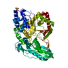
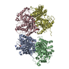
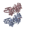
L-LEUCINE (LEU): L-LEUCINE IS BOUND TO THE SUBSTRATE BINDING SITE OF THE A DOMAIN OF SRFA-C.
Sequence details
PLEASE NOTE THAT THE GENBANK ENTRY CONTAINS THE SEQUENCE OF B. SUBTILIS STRAIN 168, WHEREAS THE ...PLEASE NOTE THAT THE GENBANK ENTRY CONTAINS THE SEQUENCE OF B. SUBTILIS STRAIN 168, WHEREAS THE RECOMBINANT PROTEIN' S SOURCE IS B. SUBTILIS STRAIN ATCC 21332. THEREFORE THERE ARE THE FOLLOWING AMINO ACID EXCHANGES COMPARED TO THE AFOREMENTIONED GENBANK ENTRY. P26A, T33S, L235P. RESIDUES 1010-1014, VPHQQ, ARE REPLACED BY ASRIKK DUE TO A FRAME SHIFT. THE PROTEIN CONTAINS A S1003A POINT MUTATION AND 29 AMINO ACIDS THAT ARE DERIVED FROM THE VECTOR'S MULTIPLE CLONING SITE, C-MYC EPITOPE SEQUENCE, A LINKER AND A HEXA- HISTIDINE TAG.
-
Experimental details
-
Experiment
Experiment
Method: X-RAY DIFFRACTION / Number of used crystals: 1
-
Sample preparation
Crystal
Density Matthews: 2.87 Å3/Da / Density % sol: 56.76 % / Description: NONE
Crystal grow
pH: 7.5 Details: 0.1 M MGCL2, 0.05 M HEPES (PH 7.5), 15 % (W/V) PEG 2000
-
Data collection
Diffraction
ID
Mean temperature (K)
Crystal-ID
1
100
1
2
1
Diffraction source
Source
Site
Beamline
ID
Wavelength
Wavelength (Å)
SYNCHROTRON
ESRF
ID14-2
1
0.933
SYNCHROTRON
SLS
X06SA
2
0.9794, 0.9792, 0.9717
Detector
Type
ID
Detector
Date
ADSC CCD
1
CCD
Jul 14, 2007
2
Radiation
ID
Protocol
Monochromatic (M) / Laue (L)
Scattering type
Wavelength-ID
1
MAD
M
x-ray
1
2
MAD
M
x-ray
1
Radiation wavelength
ID
Wavelength (Å)
Relative weight
1
0.933
1
2
0.9794
1
3
0.9792
1
4
0.9717
1
Reflection
Resolution: 2.6→82.5 Å / Num. obs: 45564 / % possible obs: 93 % / Observed criterion σ(I): -3 / Redundancy: 2.2 % / Biso Wilson estimate: 79.2 Å2 / Rmerge(I) obs: 0.04 / Net I/σ(I): 12.9
Reflection shell
Resolution: 2.6→2.67 Å / Rmerge(I) obs: 0.48 / Mean I/σ(I) obs: 2 / % possible all: 68
-
Processing
Software
Name
Version
Classification
REFMAC
5.2.0019
refinement
MOSFLM
datareduction
SCALA
datascaling
PHASER
phasing
Refinement
Method to determine structure: MAD Starting model: PDB ENTRIES 1AMU AND 2JGP
Resolution: 2.6→82.5 Å / Cor.coef. Fo:Fc: 0.949 / Cor.coef. Fo:Fc free: 0.921 / SU B: 28.59 / SU ML: 0.284 / TLS residual ADP flag: LIKELY RESIDUAL / Cross valid method: THROUGHOUT / ESU R: 0.907 / ESU R Free: 0.349 / Stereochemistry target values: MAXIMUM LIKELIHOOD Details: HYDROGENS HAVE BEEN ADDED IN THE RIDING POSITIONS. RESIDUES 1156-1160, 1175-1181 AND 1202-1206 ARE DISORDERED AND MISSING IN THE SRFA-C STRUCTURE.
Rfactor
Num. reflection
% reflection
Selection details
Rfree
0.272
1040
2.3 %
RANDOM
Rwork
0.213
-
-
-
obs
0.215
44479
93.1 %
-
Solvent computation
Ion probe radii: 0.8 Å / Shrinkage radii: 0.8 Å / VDW probe radii: 1.2 Å / Solvent model: MASK
 Movie
Movie Controller

 Yorodumi
 Open data
 Basic information
 Components
 Keywords
 Function and homology information
 X-RAY DIFFRACTION /
  Authors
 Citation
 Structure visualization
 Downloads & links
 Other downloads
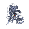


 PDBj
PDBj


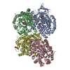
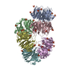
 Assembly

 Type: L-peptide linking / Mass: 131.173 Da / Num. of mol.: 1 / Source method: obtained synthetically / Formula: C6H13NO2
 Mass: 96.063 Da / Num. of mol.: 1 / Source method: obtained synthetically / Formula: SO4
 Mass: 18.015 Da / Num. of mol.: 39 / Source method: isolated from a natural source / Formula: H2O
 Sample preparation

 Processing