 Movie
Movie Controller

[English] 日本語
 Yorodumi
- PDB-3knt: Crystal structure of Methanocaldococcus jannaschii 8-oxoguanine g... -
+ Open data

- Basic information
| Entry | Database: PDB / ID: 3knt | ||||||
|---|---|---|---|---|---|---|---|
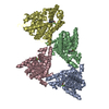
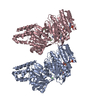
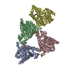
| Title | Crystal structure of Methanocaldococcus jannaschii 8-oxoguanine glycosylase/lyase in complex with 15mer DNA containing 8-oxoguanine | ||||||
 Components Components |
| ||||||
 Keywords Keywords | HYDROLASE / LYASE/DNA / protein-DNA complex / OGG / HELIX-HAIRPIN-HELIX / GLYCOSYLASE / 8-OXOGUANINE / 8-OXOG / DNA repair / DNA damage / Glycosidase / Multifunctional enzyme / Nuclease / LYASE-DNA complex | ||||||
| Function / homology |  Function and homology information Function and homology informationhydrolase activity, hydrolyzing N-glycosyl compounds / Hydrolases; Glycosylases; Hydrolysing N-glycosyl compounds / class I DNA-(apurinic or apyrimidinic site) endonuclease activity / DNA-(apurinic or apyrimidinic site) lyase / base-excision repair Similarity search - Function | ||||||
| Biological species |   Methanocaldococcus jannaschii (archaea) Methanocaldococcus jannaschii (archaea) | ||||||
| Method |  X-RAY DIFFRACTION / SYNCHROTRON / MOLECULAR REPLACEMENT / Resolution: 2.7 Å X-RAY DIFFRACTION / SYNCHROTRON / MOLECULAR REPLACEMENT / Resolution: 2.7 Å | ||||||
 Authors Authors | Faucher, F. / Doublie, S. | ||||||
 Citation Citation | Journal: J.Mol.Biol. / Year: 2010 Title: The C-terminal Lysine of Ogg2 DNA Glycosylases is a Major Molecular Determinant for Guanine/8-Oxoguanine Distinction. Authors: Faucher, F. / Wallace, S.S. / Doublie, S. | ||||||
| History |
|
- Structure visualization
| Structure viewer | Molecule: MolmilJmol/JSmol |
|---|
- Downloads & links
-Download
| PDBx/mmCIF format | 3knt.cif.gz | 242.3 KB | Display | PDBx/mmCIF format |
|---|---|---|---|---|
| PDB format | pdb3knt.ent.gz | 190.6 KB | Display | PDB format |
| PDBx/mmJSON format | 3knt.json.gz | Tree view | PDBx/mmJSON format | |
| Others |  Other downloads Other downloads |
-Validation report
| Arichive directory | https://data.pdbj.org/pub/pdb/validation_reports/kn/3kntftp://data.pdbj.org/pub/pdb/validation_reports/kn/3knt | HTTPS FTP |
|---|
-Related structure data
| Related structure data | 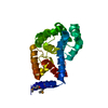 3fhfS S: Starting model for refinement |
|---|---|
| Similar structure data |


-Links
 PDBj
PDBj


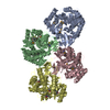
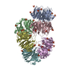
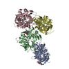
- Assembly
| Deposited unit | 
| ||||||||
|---|---|---|---|---|---|---|---|---|---|
| 1 |
| ||||||||
| 2 | 
| ||||||||
| 3 | 
| ||||||||
| 4 | 
| ||||||||
| 5 | 
| ||||||||
| Unit cell |
|
-Components
| #1: Protein | Mass: 24868.801 Da / Num. of mol.: 4 / Fragment: MjaOGG / Mutation: K129Q Source method: isolated from a genetically manipulated source Source: (gene. exp.) Methanocaldococcus jannaschii (archaea)Gene: 1451601, MJ0724, ogg / Plasmid: pET-22 / Production host:  References: UniProt: Q58134, Hydrolases; Glycosylases; Hydrolysing N-glycosyl compounds, DNA-(apurinic or apyrimidinic site) lyase #2: DNA chain | Mass: 4545.949 Da / Num. of mol.: 4 / Source method: obtained synthetically / Details: This sequence occurs naturally in humans. #3: DNA chain | Mass: 4650.021 Da / Num. of mol.: 4 / Source method: obtained synthetically / Details: This sequence occurs naturally in humans. #4: Chemical | ChemComp-NA /   Mass: 22.990 Da / Num. of mol.: 4 / Source method: obtained synthetically / Formula: Na Mass: 22.990 Da / Num. of mol.: 4 / Source method: obtained synthetically / Formula: Na#5: Water | ChemComp-HOH / |  Mass: 18.015 Da / Num. of mol.: 35 / Source method: isolated from a natural source / Formula: H2O Mass: 18.015 Da / Num. of mol.: 35 / Source method: isolated from a natural source / Formula: H2O |
|---|
-Experimental details
-Experiment
| Experiment | Method: X-RAY DIFFRACTION / Number of used crystals: 1 |
|---|
- Sample preparation
| Crystal | Density Matthews: 2.6 Å3/Da / Density % sol: 52.64 % |
|---|---|
| Crystal grow | Temperature: 277 K / Method: vapor diffusion, hanging drop / pH: 6.5 Details: PEG-8000 16%, 0.1M NaCl, pH 6.5, VAPOR DIFFUSION, HANGING DROP, temperature 277K |
-Data collection
| Diffraction | Mean temperature: 100 K |
|---|---|
| Diffraction source | Source: SYNCHROTRON / Site: APS  / Beamline: 23-ID-B / Wavelength: 1.0332 Å / Beamline: 23-ID-B / Wavelength: 1.0332 Å |
| Detector | Type: MARMOSAIC 300 mm CCD / Detector: CCD / Date: Sep 15, 2009 / Details: ADJUSTABLE FOCUS K-B PAIR SI PLUS PT, RH COATINGS |
| Radiation | Monochromator: DOUBLE CRYSTAL CRYO-COOLED SI(111) / Protocol: SINGLE WAVELENGTH / Monochromatic (M) / Laue (L): M / Scattering type: x-ray |
| Radiation wavelength | Wavelength: 1.0332 Å / Relative weight: 1 |
| Reflection | Resolution: 2.7→19.8 Å / Num. all: 38203 / Num. obs: 37810 / % possible obs: 99 % / Observed criterion σ(F): 0 / Observed criterion σ(I): 0 / Redundancy: 3.82 % / Rmerge(I) obs: 0.124 / Rsym value: 0.071 / Net I/σ(I): 12.58 |
| Reflection shell | Resolution: 2.7→2.8 Å / Redundancy: 3.87 % / Rmerge(I) obs: 0.01044 / Mean I/σ(I) obs: 1.72 / Num. unique all: 14919 / Rsym value: 0.817 / % possible all: 99.2 |
- Processing
| Software |
| ||||||||||||||||||||||||||||||||||||||||||||||||||||||||||||||||||||||||||||||||||||||||||||||||||||||||||||||||||||||||||||||||||||||||||||
|---|---|---|---|---|---|---|---|---|---|---|---|---|---|---|---|---|---|---|---|---|---|---|---|---|---|---|---|---|---|---|---|---|---|---|---|---|---|---|---|---|---|---|---|---|---|---|---|---|---|---|---|---|---|---|---|---|---|---|---|---|---|---|---|---|---|---|---|---|---|---|---|---|---|---|---|---|---|---|---|---|---|---|---|---|---|---|---|---|---|---|---|---|---|---|---|---|---|---|---|---|---|---|---|---|---|---|---|---|---|---|---|---|---|---|---|---|---|---|---|---|---|---|---|---|---|---|---|---|---|---|---|---|---|---|---|---|---|---|---|---|---|
| Refinement | Method to determine structure: MOLECULAR REPLACEMENT Starting model: 3FHF Resolution: 2.7→19.8 Å / σ(F): 2 / Stereochemistry target values: TWIN_LSQ_F
| ||||||||||||||||||||||||||||||||||||||||||||||||||||||||||||||||||||||||||||||||||||||||||||||||||||||||||||||||||||||||||||||||||||||||||||
| Solvent computation | Shrinkage radii: 0.9 Å / VDW probe radii: 1.11 Å / Solvent model: FLAT BULK SOLVENT MODEL / Bsol: 59.08 Å2 / ksol: 0.309 e/Å3 | ||||||||||||||||||||||||||||||||||||||||||||||||||||||||||||||||||||||||||||||||||||||||||||||||||||||||||||||||||||||||||||||||||||||||||||
| Refinement step | Cycle: LAST / Resolution: 2.7→19.8 Å
| ||||||||||||||||||||||||||||||||||||||||||||||||||||||||||||||||||||||||||||||||||||||||||||||||||||||||||||||||||||||||||||||||||||||||||||
| Refine LS restraints |
| ||||||||||||||||||||||||||||||||||||||||||||||||||||||||||||||||||||||||||||||||||||||||||||||||||||||||||||||||||||||||||||||||||||||||||||
| LS refinement shell |
|
