 Movie
Movie Controller

[English] 日本語
 Yorodumi
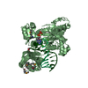
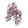
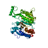
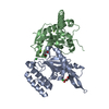
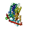
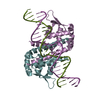
- PDB-1amu: PHENYLALANINE ACTIVATING DOMAIN OF GRAMICIDIN SYNTHETASE 1 IN A C... -
+ Open data

- Basic information
| Entry | Database: PDB / ID: 1amu | ||||||
|---|---|---|---|---|---|---|---|
| Title | PHENYLALANINE ACTIVATING DOMAIN OF GRAMICIDIN SYNTHETASE 1 IN A COMPLEX WITH AMP AND PHENYLALANINE | ||||||
 Components Components | GRAMICIDIN SYNTHETASE 1 | ||||||
 Keywords Keywords | PEPTIDE SYNTHETASE / GRSA / ADENYLATE FORMING | ||||||
| Function / homology |  Function and homology information Function and homology informationphenylalanine racemase (ATP-hydrolysing) / phenylalanine racemase (ATP-hydrolyzing) activity / amino acid activation for nonribosomal peptide biosynthetic process / secondary metabolite biosynthetic process / lipid biosynthetic process / ligase activity / antibiotic biosynthetic process / ATP binding Similarity search - Function | ||||||
| Biological species |  Brevibacillus brevis (bacteria) Brevibacillus brevis (bacteria) | ||||||
| Method |  X-RAY DIFFRACTION / SYNCHROTRON / MIR / Resolution: 1.9 Å X-RAY DIFFRACTION / SYNCHROTRON / MIR / Resolution: 1.9 Å | ||||||
 Authors Authors | Conti, E. / Stachelhaus, T. / Marahiel, M.A. / Brick, P. | ||||||
 Citation Citation | Journal: EMBO J. / Year: 1997 Title: Structural basis for the activation of phenylalanine in the non-ribosomal biosynthesis of gramicidin S. Authors: Conti, E. / Stachelhaus, T. / Marahiel, M.A. / Brick, P. #1: Journal: J.Biol.Chem. / Year: 1995Title: Modular Structure of Peptide Synthetases Revealed by Dissection of the Multifunctional Enzyme Grsa Authors: Stachelhaus, T. / Marahiel, M.A. | ||||||
| History |
|
- Structure visualization
| Structure viewer | Molecule: MolmilJmol/JSmol |
|---|
- Downloads & links
-Download
| PDBx/mmCIF format | 1amu.cif.gz | 225.3 KB | Display | PDBx/mmCIF format |
|---|---|---|---|---|
| PDB format | pdb1amu.ent.gz | 176.7 KB | Display | PDB format |
| PDBx/mmJSON format | 1amu.json.gz | Tree view | PDBx/mmJSON format | |
| Others |  Other downloads Other downloads |
-Validation report
| Arichive directory | https://data.pdbj.org/pub/pdb/validation_reports/am/1amuftp://data.pdbj.org/pub/pdb/validation_reports/am/1amu | HTTPS FTP |
|---|
-Related structure data
| Similar structure data |
|---|


-Links
 PDBj
PDBj


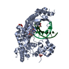
- Assembly
| Deposited unit | 
| ||||||||
|---|---|---|---|---|---|---|---|---|---|
| 1 | 
| ||||||||
| 2 | 
| ||||||||
| Unit cell |
|
-Components
-Protein , 1 types, 2 molecules AB
| #1: Protein | Mass: 64131.793 Da / Num. of mol.: 2 Fragment: ADENYLATE FORMING DOMAIN, RESIDUES 1 - 556 PLUS A 7 RESIDUE C-TERMINAL TAG - SHHHHHH Source method: isolated from a genetically manipulated source Source: (gene. exp.) Brevibacillus brevis (bacteria) / Gene: GRSA / Plasmid: PQE60 / Production host: References: UniProt: P14687, UniProt: P0C061*PLUS, phenylalanine racemase (ATP-hydrolysing) |
|---|
-Non-polymers , 5 types, 577 molecules 


| #2: Chemical |  Mass: 24.305 Da / Num. of mol.: 2 / Source method: obtained synthetically / Formula: Mg Mass: 24.305 Da / Num. of mol.: 2 / Source method: obtained synthetically / Formula: Mg#3: Chemical |  Mass: 96.063 Da / Num. of mol.: 2 / Source method: obtained synthetically / Formula: SO4 Mass: 96.063 Da / Num. of mol.: 2 / Source method: obtained synthetically / Formula: SO4#4: Chemical |  Type: L-peptide linking / Mass: 165.189 Da / Num. of mol.: 2 / Source method: obtained synthetically / Formula: C9H11NO2 Type: L-peptide linking / Mass: 165.189 Da / Num. of mol.: 2 / Source method: obtained synthetically / Formula: C9H11NO2#5: Chemical |  Mass: 347.221 Da / Num. of mol.: 2 / Source method: obtained synthetically / Formula: C10H14N5O7P / Comment: AMP*YM Mass: 347.221 Da / Num. of mol.: 2 / Source method: obtained synthetically / Formula: C10H14N5O7P / Comment: AMP*YM#6: Water | ChemComp-HOH / | Mass: 18.015 Da / Num. of mol.: 569 / Source method: isolated from a natural source / Formula: H2O |
|---|
-Experimental details
-Experiment
| Experiment | Method: X-RAY DIFFRACTION / Number of used crystals: 1 |
|---|
- Sample preparation
| Crystal | Density Matthews: 2.5 Å3/Da / Density % sol: 50 % | ||||||||||||||||||||||||||||||||||||||||||||||||||||||||||||||||||
|---|---|---|---|---|---|---|---|---|---|---|---|---|---|---|---|---|---|---|---|---|---|---|---|---|---|---|---|---|---|---|---|---|---|---|---|---|---|---|---|---|---|---|---|---|---|---|---|---|---|---|---|---|---|---|---|---|---|---|---|---|---|---|---|---|---|---|---|
| Crystal grow | Temperature: 291 K / Method: vapor diffusion / pH: 6.5 Details: CRYSTALS WERE GROWN USING THE VAPOR DIFFUSION TECHNIQUE. THE PROTEIN AT 15 MG/ML IN 10 MM TRIS-HCL PH7.8, 50 MM NACL, 15% GLYCEROL, 2 MM ATP, 2 MM L-PHE AND 4 MM MGCL2 WAS EQUILIBRATED WITH ...Details: CRYSTALS WERE GROWN USING THE VAPOR DIFFUSION TECHNIQUE. THE PROTEIN AT 15 MG/ML IN 10 MM TRIS-HCL PH7.8, 50 MM NACL, 15% GLYCEROL, 2 MM ATP, 2 MM L-PHE AND 4 MM MGCL2 WAS EQUILIBRATED WITH AN EQUAL VOLUME OF RESERVOIR SOLUTION CONTAINING 28-32%(W/V) METHOXY POLYETHYLENE GLYCOL 5000, 200 MM AMMONIUM SULFATE, AND 100 MM ADA PH6.5 AT 18C., vapor diffusion, temperature 291K | ||||||||||||||||||||||||||||||||||||||||||||||||||||||||||||||||||
| Crystal grow | *PLUS Temperature: 18 ℃ / pH: 7.8 / Method: vapor diffusion, hanging drop | ||||||||||||||||||||||||||||||||||||||||||||||||||||||||||||||||||
| Components of the solutions | *PLUS
|
-Data collection
| Diffraction | Mean temperature: 100 K |
|---|---|
| Diffraction source | Source: SYNCHROTRON / Site: EMBL/DESY, HAMBURG  / Beamline: BW7B / Wavelength: 0.88 / Beamline: BW7B / Wavelength: 0.88 |
| Detector | Type: MAR scanner 300 mm plate / Detector: IMAGE PLATE / Date: Sep 9, 1996 |
| Radiation | Monochromator: Y / Monochromatic (M) / Laue (L): M / Scattering type: x-ray |
| Radiation wavelength | Wavelength: 0.88 Å / Relative weight: 1 |
| Reflection | Resolution: 1.9→20 Å / Num. obs: 91144 / % possible obs: 96.4 % / Redundancy: 2.5 % / Rmerge(I) obs: 0.051 / Net I/σ(I): 7.3 |
| Reflection shell | Resolution: 1.9→2 Å / Redundancy: 1.9 % / Rmerge(I) obs: 0.232 / Mean I/σ(I) obs: 2.7 / % possible all: 88.3 |
| Reflection | *PLUS Num. measured all: 230285 |
| Reflection shell | *PLUS % possible obs: 88.3 % |
- Processing
| Software |
| ||||||||||||||||||||||||||||||||||||||||||||||||||||||||||||||||||||||||||||||||
|---|---|---|---|---|---|---|---|---|---|---|---|---|---|---|---|---|---|---|---|---|---|---|---|---|---|---|---|---|---|---|---|---|---|---|---|---|---|---|---|---|---|---|---|---|---|---|---|---|---|---|---|---|---|---|---|---|---|---|---|---|---|---|---|---|---|---|---|---|---|---|---|---|---|---|---|---|---|---|---|---|---|
| Refinement | Method to determine structure: MIR / Resolution: 1.9→20 Å / Rfactor Rfree error: 0.004 / Data cutoff high absF: 0 / Data cutoff low absF: 0 / Isotropic thermal model: RESTRAINED / Cross valid method: THROUGHOUT / σ(F): 0
| ||||||||||||||||||||||||||||||||||||||||||||||||||||||||||||||||||||||||||||||||
| Displacement parameters | Biso mean: 33 Å2 | ||||||||||||||||||||||||||||||||||||||||||||||||||||||||||||||||||||||||||||||||
| Refinement step | Cycle: LAST / Resolution: 1.9→20 Å
| ||||||||||||||||||||||||||||||||||||||||||||||||||||||||||||||||||||||||||||||||
| Refine LS restraints |
| ||||||||||||||||||||||||||||||||||||||||||||||||||||||||||||||||||||||||||||||||
| Refine LS restraints NCS | NCS model details: UNRESTRAINED | ||||||||||||||||||||||||||||||||||||||||||||||||||||||||||||||||||||||||||||||||
| LS refinement shell | Resolution: 1.9→1.99 Å / Rfactor Rfree error: 0.016 / Total num. of bins used: 8
| ||||||||||||||||||||||||||||||||||||||||||||||||||||||||||||||||||||||||||||||||
| Xplor file |
| ||||||||||||||||||||||||||||||||||||||||||||||||||||||||||||||||||||||||||||||||
| Software | *PLUS Name: X-PLOR / Version: 3.1 / Classification: refinement | ||||||||||||||||||||||||||||||||||||||||||||||||||||||||||||||||||||||||||||||||
| Refine LS restraints | *PLUS
|
