 Movie
Movie Controller

[English] 日本語
 Yorodumi
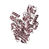
- PDB-5brq: Crystal structure of Bacillus licheniformis trehalose-6-phosphate... -
+ Open data

- Basic information
| Entry | Database: PDB / ID: 5brq | ||||||
|---|---|---|---|---|---|---|---|
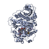
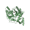
| Title | Crystal structure of Bacillus licheniformis trehalose-6-phosphate hydrolase (TreA) | ||||||
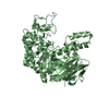
 Components Components | Glycoside Hydrolase Family 13 | ||||||
 Keywords Keywords | HYDROLASE / trehalose-6-phosphate hydrolase / TIM barrel / GH13 family | ||||||
| Function / homology |  Function and homology information Function and homology informationalpha,alpha-phosphotrehalase / alpha,alpha-phosphotrehalase activity / trehalose catabolic process / alpha-amylase activity / metal ion binding / cytoplasm Similarity search - Function | ||||||
| Biological species |  | ||||||
| Method |  X-RAY DIFFRACTION / SYNCHROTRON / MOLECULAR REPLACEMENT / Resolution: 2.003 Å X-RAY DIFFRACTION / SYNCHROTRON / MOLECULAR REPLACEMENT / Resolution: 2.003 Å | ||||||
 Authors Authors | Hsiao, C.-D. / Lin, M.-G. | ||||||
 Citation Citation | Journal: Acta Crystallogr D Struct Biol / Year: 2016 Title: Bacillus licheniformis trehalose-6-phosphate hydrolase structures suggest keys to substrate specificity Authors: Lin, M.-G. / Chi, M.-C. / Naveen, V. / Li, Y.-C. / Lin, L.-L. / Hsiao, C.-D. | ||||||
| History |
|
- Structure visualization
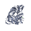
| Structure viewer | Molecule: MolmilJmol/JSmol |
|---|
- Downloads & links
-Download
| PDBx/mmCIF format | 5brq.cif.gz | 488.6 KB | Display | PDBx/mmCIF format |
|---|---|---|---|---|
| PDB format | pdb5brq.ent.gz | 396.9 KB | Display | PDB format |
| PDBx/mmJSON format | 5brq.json.gz | Tree view | PDBx/mmJSON format | |
| Others |  Other downloads Other downloads |
-Validation report
| Arichive directory | https://data.pdbj.org/pub/pdb/validation_reports/br/5brqftp://data.pdbj.org/pub/pdb/validation_reports/br/5brq | HTTPS FTP |
|---|
-Related structure data
| Related structure data |  5brpC  1uokS C: citing same article ( S: Starting model for refinement |
|---|---|
| Similar structure data |
-Links
 PDBj
PDBj

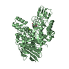
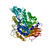
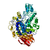
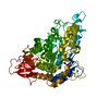
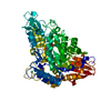
- Assembly
| Deposited unit | 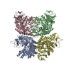
| ||||||||
|---|---|---|---|---|---|---|---|---|---|
| 1 | 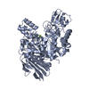
| ||||||||
| 2 | 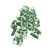
| ||||||||
| 3 | 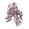
| ||||||||
| 4 | 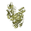
| ||||||||
| Unit cell |
|
-Components
| #1: Protein | Mass: 66642.312 Da / Num. of mol.: 4 Source method: isolated from a genetically manipulated source Source: (gene. exp.) Strain: ATCC 14580 = DSM 13 / Gene: treA, BL03069 / Plasmid: pQE30 / Production host: #2: Chemical | ChemComp-MG /   Mass: 24.305 Da / Num. of mol.: 4 / Source method: obtained synthetically / Formula: Mg Mass: 24.305 Da / Num. of mol.: 4 / Source method: obtained synthetically / Formula: Mg#3: Water | ChemComp-HOH / |  Mass: 18.015 Da / Num. of mol.: 1445 / Source method: isolated from a natural source / Formula: H2O Mass: 18.015 Da / Num. of mol.: 1445 / Source method: isolated from a natural source / Formula: H2O |
|---|
-Experimental details
-Experiment
| Experiment | Method: X-RAY DIFFRACTION / Number of used crystals: 1 |
|---|
- Sample preparation
| Crystal | Density Matthews: 2.25 Å3/Da / Density % sol: 45.4 % |
|---|---|
| Crystal grow | Temperature: 298 K / Method: vapor diffusion, hanging drop / pH: 5 Details: 20% (w/v) PEG 3350, 0.2 M magnesium acetate hexahydrate, 2% Tacsimate (pH 5.0) |
-Data collection
| Diffraction | Mean temperature: 100 K |
|---|---|
| Diffraction source | Source: SYNCHROTRON / Site: NSRRC  / Beamline: BL13B1 / Wavelength: 0.903 Å / Beamline: BL13B1 / Wavelength: 0.903 Å |
| Detector | Type: ADSC QUANTUM 315r / Detector: CCD / Date: Nov 5, 2013 |
| Radiation | Protocol: SINGLE WAVELENGTH / Monochromatic (M) / Laue (L): M / Scattering type: x-ray |
| Radiation wavelength | Wavelength: 0.903 Å / Relative weight: 1 |
| Reflection | Resolution: 2→30 Å / Num. obs: 149079 / % possible obs: 96.8 % / Redundancy: 3.8 % / Net I/σ(I): 29 |
- Processing
| Software | Name: PHENIX / Version: 1.8.1_1168 / Classification: refinement | |||||||||||||||||||||||||||||||||||||||||||||||||||||||||||||||||||||||||||||||||||||||||||||||||||||||||
|---|---|---|---|---|---|---|---|---|---|---|---|---|---|---|---|---|---|---|---|---|---|---|---|---|---|---|---|---|---|---|---|---|---|---|---|---|---|---|---|---|---|---|---|---|---|---|---|---|---|---|---|---|---|---|---|---|---|---|---|---|---|---|---|---|---|---|---|---|---|---|---|---|---|---|---|---|---|---|---|---|---|---|---|---|---|---|---|---|---|---|---|---|---|---|---|---|---|---|---|---|---|---|---|---|---|---|
| Refinement | Method to determine structure: MOLECULAR REPLACEMENT Starting model: 1UOK Resolution: 2.003→24.304 Å / SU ML: 0.26 / Cross valid method: FREE R-VALUE / σ(F): 1.97 / Phase error: 25.73 / Stereochemistry target values: ML
| |||||||||||||||||||||||||||||||||||||||||||||||||||||||||||||||||||||||||||||||||||||||||||||||||||||||||
| Solvent computation | Shrinkage radii: 0.9 Å / VDW probe radii: 1.11 Å / Solvent model: FLAT BULK SOLVENT MODEL | |||||||||||||||||||||||||||||||||||||||||||||||||||||||||||||||||||||||||||||||||||||||||||||||||||||||||
| Refinement step | Cycle: LAST / Resolution: 2.003→24.304 Å
| |||||||||||||||||||||||||||||||||||||||||||||||||||||||||||||||||||||||||||||||||||||||||||||||||||||||||
| Refine LS restraints |
| |||||||||||||||||||||||||||||||||||||||||||||||||||||||||||||||||||||||||||||||||||||||||||||||||||||||||
| LS refinement shell |
|
