 Movie
Movie Controller

[English] 日本語
 Yorodumi
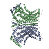
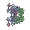
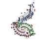
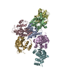
- PDB-4bxs: Crystal Structure of the Prothrombinase Complex from the Venom of... -
+ Open data

- Basic information
| Entry | Database: PDB / ID: 4bxs | |||||||||
|---|---|---|---|---|---|---|---|---|---|---|
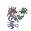
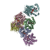
| Title | Crystal Structure of the Prothrombinase Complex from the Venom of Pseudonaja Textilis | |||||||||
 Components Components |
| |||||||||
 Keywords Keywords | BLOOD CLOTTING / BLOOD COAGULATION / PROTHROMBINASE / HYDROLASE | |||||||||
| Function / homology |  Function and homology information Function and homology informationvenom-mediated blood coagulation / coagulation factor Xa / peptidase activator activity / blood coagulation / toxin activity / signaling receptor activity / copper ion binding / serine-type endopeptidase activity / calcium ion binding / protein-containing complex ...venom-mediated blood coagulation / coagulation factor Xa / peptidase activator activity / blood coagulation / toxin activity / signaling receptor activity / copper ion binding / serine-type endopeptidase activity / calcium ion binding / protein-containing complex / proteolysis / : / extracellular region / plasma membrane Similarity search - Function | |||||||||
| Biological species |  PSEUDONAJA TEXTILIS (eastern brown snake) PSEUDONAJA TEXTILIS (eastern brown snake) | |||||||||
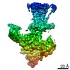
| Method |  X-RAY DIFFRACTION / SYNCHROTRON / MOLECULAR REPLACEMENT / Resolution: 3.32 Å X-RAY DIFFRACTION / SYNCHROTRON / MOLECULAR REPLACEMENT / Resolution: 3.32 Å | |||||||||
 Authors Authors | Lechtenberg, B.C. / Murray-Rust, T.A. / Johnson, D.J.D. / Adams, T.E. / Krishnaswamy, S. / Camire, R.M. / Huntington, J.A. | |||||||||
 Citation Citation | Journal: Blood / Year: 2013 Title: Crystal Structure of the Prothrombinase Complex from the Venom of Pseudonaja Textilis. Authors: Lechtenberg, B.C. / Murray-Rust, T.A. / Johnson, D.J. / Adams, T.E. / Krishnaswamy, S. / Camire, R.M. / Huntington, J.A. | |||||||||
| History |
|
- Structure visualization
| Structure viewer | Molecule: MolmilJmol/JSmol |
|---|
- Downloads & links
-Download
| PDBx/mmCIF format | 4bxs.cif.gz | 322.6 KB | Display | PDBx/mmCIF format |
|---|---|---|---|---|
| PDB format | pdb4bxs.ent.gz | 242.7 KB | Display | PDB format |
| PDBx/mmJSON format | 4bxs.json.gz | Tree view | PDBx/mmJSON format | |
| Others |  Other downloads Other downloads |
-Validation report
| Arichive directory | https://data.pdbj.org/pub/pdb/validation_reports/bx/4bxsftp://data.pdbj.org/pub/pdb/validation_reports/bx/4bxs | HTTPS FTP |
|---|
-Related structure data
| Related structure data |  4bxwC  1kigS C: citing same article ( S: Starting model for refinement |
|---|---|
| Similar structure data |
-Links
 PDBj
PDBj


- Assembly
| Deposited unit | 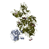
| ||||||||
|---|---|---|---|---|---|---|---|---|---|
| 1 |
| ||||||||
| Unit cell |
|
-Components
-Protein , 2 types, 2 molecules AV
| #1: Protein | Mass: 47041.273 Da / Num. of mol.: 1 / Fragment: EGF2-CATALYTIC DOMAIN CONSTRUCT Source method: isolated from a genetically manipulated source Details: PSEUTARIN C CATALYTIC SUBUNIT Source: (gene. exp.) PSEUDONAJA TEXTILIS (eastern brown snake)Organ: VENOM GLAND / Production host:  |
|---|---|
| #2: Protein | Mass: 162826.781 Da / Num. of mol.: 1 Source method: isolated from a genetically manipulated source Source: (gene. exp.) PSEUDONAJA TEXTILIS (eastern brown snake)Organ: VENOM GLAND / Plasmid: PED / Cell line (production host): BHK-M / Production host:   CRICETULUS GRISEUS (Chinese hamster) / References: UniProt: Q7SZN0 CRICETULUS GRISEUS (Chinese hamster) / References: UniProt: Q7SZN0 |
-Sugars , 4 types, 5 molecules 
| #3: Polysaccharide | alpha-L-fucopyranose-(1-6)-2-acetamido-2-deoxy-beta-D-glucopyranose Source method: isolated from a genetically manipulated source | ||
|---|---|---|---|
| #4: Polysaccharide | alpha-D-mannopyranose-(1-2)-alpha-D-mannopyranose-(1-3)-[alpha-D-mannopyranose-(1-3)-[alpha-D- ...alpha-D-mannopyranose-(1-2)-alpha-D-mannopyranose-(1-3)-[alpha-D-mannopyranose-(1-3)-[alpha-D-mannopyranose-(1-6)]alpha-D-mannopyranose-(1-6)]beta-D-mannopyranose-(1-4)-2-acetamido-2-deoxy-beta-D-glucopyranose-(1-4)-2-acetamido-2-deoxy-beta-D-glucopyranose Source method: isolated from a genetically manipulated source | ||
| #5: Polysaccharide | Source method: isolated from a genetically manipulated source #6: Sugar | ChemComp-NAG / |  Type: D-saccharide, beta linking / Mass: 221.208 Da / Num. of mol.: 1 Type: D-saccharide, beta linking / Mass: 221.208 Da / Num. of mol.: 1Source method: isolated from a genetically manipulated source Formula: C8H15NO6 |
-Non-polymers , 3 types, 115 molecules 


| #7: Chemical |  Mass: 40.078 Da / Num. of mol.: 2 / Source method: obtained synthetically / Formula: Ca Mass: 40.078 Da / Num. of mol.: 2 / Source method: obtained synthetically / Formula: Ca#8: Chemical | ChemComp-CU / |  Mass: 63.546 Da / Num. of mol.: 1 / Source method: obtained synthetically / Formula: Cu Mass: 63.546 Da / Num. of mol.: 1 / Source method: obtained synthetically / Formula: Cu#9: Water | ChemComp-HOH / | Mass: 18.015 Da / Num. of mol.: 112 / Source method: isolated from a natural source / Formula: H2O |
|---|
-Details
| Has protein modification | Y |
|---|
-Experimental details
-Experiment
| Experiment | Method: X-RAY DIFFRACTION / Number of used crystals: 1 |
|---|
- Sample preparation
| Crystal | Density Matthews: 3.72 Å3/Da / Density % sol: 66.98 % / Description: NONE |
|---|---|
| Crystal grow | pH: 8 Details: 0.1 M MORPHEUS BUFFER 3, 20% MORPHEUS P20K_P550MME, 50 MM MORPHEUS CARBOXYLIC ACIDS, pH 8 |
-Data collection
| Diffraction | Mean temperature: 100 K |
|---|---|
| Diffraction source | Source: SYNCHROTRON / Site: Diamond  / Beamline: I04 / Wavelength: 0.8856 / Beamline: I04 / Wavelength: 0.8856 |
| Detector | Type: ADSC CCD / Detector: CCD / Date: Jul 21, 2012 |
| Radiation | Protocol: SINGLE WAVELENGTH / Monochromatic (M) / Laue (L): M / Scattering type: x-ray |
| Radiation wavelength | Wavelength: 0.8856 Å / Relative weight: 1 |
| Reflection | Resolution: 3.32→101.59 Å / Num. obs: 43913 / % possible obs: 99.4 % / Observed criterion σ(I): 0 / Redundancy: 6 % / Rmerge(I) obs: 0.15 / Net I/σ(I): 7.8 |
| Reflection shell | Resolution: 3.32→3.5 Å / Redundancy: 5.4 % / Rmerge(I) obs: 1 / Mean I/σ(I) obs: 1.5 / % possible all: 95.1 |
- Processing
| Software |
| ||||||||||||||||||||||||||||||||||||||||||||||||||||||||||||||||||||||||||||||||||||||||||||||||||||||||||||||||||||||||||||||||||||||||||||||||||||||||||||||||||||||||||||||||||||||
|---|---|---|---|---|---|---|---|---|---|---|---|---|---|---|---|---|---|---|---|---|---|---|---|---|---|---|---|---|---|---|---|---|---|---|---|---|---|---|---|---|---|---|---|---|---|---|---|---|---|---|---|---|---|---|---|---|---|---|---|---|---|---|---|---|---|---|---|---|---|---|---|---|---|---|---|---|---|---|---|---|---|---|---|---|---|---|---|---|---|---|---|---|---|---|---|---|---|---|---|---|---|---|---|---|---|---|---|---|---|---|---|---|---|---|---|---|---|---|---|---|---|---|---|---|---|---|---|---|---|---|---|---|---|---|---|---|---|---|---|---|---|---|---|---|---|---|---|---|---|---|---|---|---|---|---|---|---|---|---|---|---|---|---|---|---|---|---|---|---|---|---|---|---|---|---|---|---|---|---|---|---|---|---|
| Refinement | Method to determine structure: MOLECULAR REPLACEMENT Starting model: UNPUBLISHED FV STRUCTURE AND PDB ENTRY 1KIG Resolution: 3.32→89.81 Å / Cor.coef. Fo:Fc: 0.891 / Cor.coef. Fo:Fc free: 0.81 / SU B: 38.223 / SU ML: 0.605 / Cross valid method: THROUGHOUT / ESU R Free: 0.648 / Stereochemistry target values: MAXIMUM LIKELIHOOD Details: HYDROGENS HAVE BEEN ADDED IN THE RIDING POSITIONS. DISORDERED SIDE CHAINS AND LOOPS WERE NOT MODELLED, WITH THE EXCEPTION OF A PORTION OF THE A2-REGION OF FV (656-670)
| ||||||||||||||||||||||||||||||||||||||||||||||||||||||||||||||||||||||||||||||||||||||||||||||||||||||||||||||||||||||||||||||||||||||||||||||||||||||||||||||||||||||||||||||||||||||
| Solvent computation | Ion probe radii: 0.8 Å / Shrinkage radii: 0.8 Å / VDW probe radii: 1.2 Å / Solvent model: MASK | ||||||||||||||||||||||||||||||||||||||||||||||||||||||||||||||||||||||||||||||||||||||||||||||||||||||||||||||||||||||||||||||||||||||||||||||||||||||||||||||||||||||||||||||||||||||
| Displacement parameters | Biso mean: 117.583 Å2
| ||||||||||||||||||||||||||||||||||||||||||||||||||||||||||||||||||||||||||||||||||||||||||||||||||||||||||||||||||||||||||||||||||||||||||||||||||||||||||||||||||||||||||||||||||||||
| Refinement step | Cycle: LAST / Resolution: 3.32→89.81 Å
| ||||||||||||||||||||||||||||||||||||||||||||||||||||||||||||||||||||||||||||||||||||||||||||||||||||||||||||||||||||||||||||||||||||||||||||||||||||||||||||||||||||||||||||||||||||||
| Refine LS restraints |
|