 Movie
Movie Controller

[English] 日本語
 Yorodumi
- PDB-7dsb: Crystal structure of actin capping protein in complex with V-1 an... -
+ Open data

- Basic information
| Entry | Database: PDB / ID: 7dsb | ||||||||||||
|---|---|---|---|---|---|---|---|---|---|---|---|---|---|
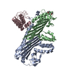
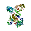
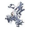
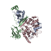
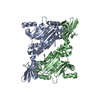
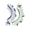
| Title | Crystal structure of actin capping protein in complex with V-1 and twinfilin C-terminal tail | ||||||||||||
 Components Components |
| ||||||||||||
 Keywords Keywords | CYTOSOLIC PROTEIN / actin dynamics / actin capping protein / twinfilin / CARMIL / V-1 | ||||||||||||
| Function / homology |  Function and homology information Function and homology informationregulation of barbed-end actin filament capping / positive regulation of macromolecule biosynthetic process / Advanced glycosylation endproduct receptor signaling / : / RHOF GTPase cycle / HSP90 chaperone cycle for steroid hormone receptors (SHR) in the presence of ligand / COPI-independent Golgi-to-ER retrograde traffic / RHOBTB2 GTPase cycle / Factors involved in megakaryocyte development and platelet production / COPI-mediated anterograde transport ...regulation of barbed-end actin filament capping / positive regulation of macromolecule biosynthetic process / Advanced glycosylation endproduct receptor signaling / : / RHOF GTPase cycle / HSP90 chaperone cycle for steroid hormone receptors (SHR) in the presence of ligand / COPI-independent Golgi-to-ER retrograde traffic / RHOBTB2 GTPase cycle / Factors involved in megakaryocyte development and platelet production / COPI-mediated anterograde transport / negative regulation of filopodium assembly / regulation of striated muscle tissue development / F-actin capping protein complex / WASH complex / negative regulation of actin filament polymerization / cell junction assembly / barbed-end actin filament capping / actin polymerization or depolymerization / actin filament depolymerization / regulation of lamellipodium assembly / regulation of cell morphogenesis / lamellipodium assembly / regulation of cell size / positive regulation of cardiac muscle hypertrophy / cortical cytoskeleton / positive regulation of protein metabolic process / myofibril / brush border / sperm head-tail coupling apparatus / actin monomer binding / phosphatidylinositol-4,5-bisphosphate binding / cytoskeleton organization / : / filopodium / actin filament / positive regulation of neuron projection development / Schaffer collateral - CA1 synapse / cell morphogenesis / Z disc / neuron differentiation / cell-cell junction / actin filament binding / regulation of translation / lamellipodium / actin cytoskeleton / positive regulation of cell growth / actin binding / actin cytoskeleton organization / protein tyrosine kinase activity / postsynaptic density / protein-containing complex binding / perinuclear region of cytoplasm / ATP binding / membrane / nucleus / plasma membrane / cytoplasm / cytosol Similarity search - Function | ||||||||||||
| Biological species |   Homo sapiens (human) Homo sapiens (human) | ||||||||||||
| Method |  X-RAY DIFFRACTION / SYNCHROTRON / MOLECULAR REPLACEMENT / molecular replacement / Resolution: 2.44 Å X-RAY DIFFRACTION / SYNCHROTRON / MOLECULAR REPLACEMENT / molecular replacement / Resolution: 2.44 Å | ||||||||||||
 Authors Authors | Takeda, S. | ||||||||||||
| Funding support |  Japan, 3items Japan, 3items
| ||||||||||||
 Citation Citation | Journal: J.Mol.Biol. / Year: 2021 Title: Structural Insights into the Regulation of Actin Capping Protein by Twinfilin C-terminal Tail. Authors: Takeda, S. / Koike, R. / Fujiwara, I. / Narita, A. / Miyata, M. / Ota, M. / Maeda, Y. | ||||||||||||
| History |
|
- Structure visualization
| Structure viewer | Molecule: MolmilJmol/JSmol |
|---|
- Downloads & links
-Download
| PDBx/mmCIF format | 7dsb.cif.gz | 154 KB | Display | PDBx/mmCIF format |
|---|---|---|---|---|
| PDB format | pdb7dsb.ent.gz | 108.4 KB | Display | PDB format |
| PDBx/mmJSON format | 7dsb.json.gz | Tree view | PDBx/mmJSON format | |
| Others |  Other downloads Other downloads |
-Validation report
| Arichive directory | https://data.pdbj.org/pub/pdb/validation_reports/ds/7dsbftp://data.pdbj.org/pub/pdb/validation_reports/ds/7dsb | HTTPS FTP |
|---|
-Related structure data
| Related structure data |  7ds2C  7ds3C  7ds4C  7ds6C 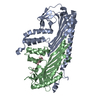 7ds8C 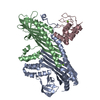 7dsaC 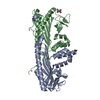 3aaaS S: Starting model for refinement C: citing same article ( |
|---|---|
| Similar structure data |

-Links
 PDBj
PDBj
- Assembly
| Deposited unit | 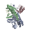
| ||||||||||||
|---|---|---|---|---|---|---|---|---|---|---|---|---|---|
| 1 |
| ||||||||||||
| Unit cell |
|
-Components
| #1: Protein | Mass: 33001.789 Da / Num. of mol.: 1 Source method: isolated from a genetically manipulated source Source: (gene. exp.)  |
|---|---|
| #2: Protein | Mass: 27473.070 Da / Num. of mol.: 1 Source method: isolated from a genetically manipulated source Source: (gene. exp.) |
| #3: Protein | Mass: 13324.238 Da / Num. of mol.: 1 Source method: isolated from a genetically manipulated source Source: (gene. exp.) Homo sapiens (human) / Gene: MTPN / Production host: |
| #4: Protein/peptide | Mass: 3331.942 Da / Num. of mol.: 1 / Source method: obtained synthetically / Source: (synth.) |
| #5: Water | ChemComp-HOH /  Mass: 18.015 Da / Num. of mol.: 94 / Source method: isolated from a natural source / Formula: H2O Mass: 18.015 Da / Num. of mol.: 94 / Source method: isolated from a natural source / Formula: H2O |
-Experimental details
-Experiment
| Experiment | Method: X-RAY DIFFRACTION / Number of used crystals: 1 |
|---|
- Sample preparation
| Crystal | Density Matthews: 2.41 Å3/Da / Density % sol: 48.95 % |
|---|---|
| Crystal grow | Temperature: 293 K / Method: vapor diffusion, hanging drop / pH: 7.5 Details: 10% (w/v) PEG 3350, 5% (v/v) isopropanol, 0.1M HEPES-NaOH (pH = 7.5) |
-Data collection
| Diffraction | Mean temperature: 90 K / Serial crystal experiment: N | ||||||||||||||||||||||||||||||||||||||||||||||||||||||||||||||||||||||||||||||||
|---|---|---|---|---|---|---|---|---|---|---|---|---|---|---|---|---|---|---|---|---|---|---|---|---|---|---|---|---|---|---|---|---|---|---|---|---|---|---|---|---|---|---|---|---|---|---|---|---|---|---|---|---|---|---|---|---|---|---|---|---|---|---|---|---|---|---|---|---|---|---|---|---|---|---|---|---|---|---|---|---|---|
| Diffraction source | Source: SYNCHROTRON / Site: AichiSR / Beamline: BL2S1 / Wavelength: 1.12 Å | ||||||||||||||||||||||||||||||||||||||||||||||||||||||||||||||||||||||||||||||||
| Detector | Type: ADSC QUANTUM 270 / Detector: CCD / Date: Nov 12, 2020 | ||||||||||||||||||||||||||||||||||||||||||||||||||||||||||||||||||||||||||||||||
| Radiation | Protocol: SINGLE WAVELENGTH / Monochromatic (M) / Laue (L): M / Scattering type: x-ray | ||||||||||||||||||||||||||||||||||||||||||||||||||||||||||||||||||||||||||||||||
| Radiation wavelength | Wavelength: 1.12 Å / Relative weight: 1 | ||||||||||||||||||||||||||||||||||||||||||||||||||||||||||||||||||||||||||||||||
| Reflection | Resolution: 2.44→45.84 Å / Num. obs: 28229 / % possible obs: 99.8 % / Redundancy: 6.009 % / Biso Wilson estimate: 50.147 Å2 / CC1/2: 0.998 / Rmerge(I) obs: 0.098 / Rrim(I) all: 0.107 / Χ2: 0.858 / Net I/σ(I): 15.84 | ||||||||||||||||||||||||||||||||||||||||||||||||||||||||||||||||||||||||||||||||
| Reflection shell | Diffraction-ID: 1
|
-Phasing
| Phasing | Method: molecular replacement |
|---|
- Processing
| Software |
| |||||||||||||||||||||||||||||||||||||||||||||||||||||||||||||||||||||||||||||
|---|---|---|---|---|---|---|---|---|---|---|---|---|---|---|---|---|---|---|---|---|---|---|---|---|---|---|---|---|---|---|---|---|---|---|---|---|---|---|---|---|---|---|---|---|---|---|---|---|---|---|---|---|---|---|---|---|---|---|---|---|---|---|---|---|---|---|---|---|---|---|---|---|---|---|---|---|---|---|
| Refinement | Method to determine structure: MOLECULAR REPLACEMENT Starting model: 3aaa Resolution: 2.44→45.84 Å / SU ML: 0.3142 / Cross valid method: FREE R-VALUE / σ(F): 1.36 / Phase error: 25.407 Stereochemistry target values: GeoStd + Monomer Library + CDL v1.2
| |||||||||||||||||||||||||||||||||||||||||||||||||||||||||||||||||||||||||||||
| Solvent computation | Shrinkage radii: 0.9 Å / VDW probe radii: 1.11 Å / Solvent model: FLAT BULK SOLVENT MODEL | |||||||||||||||||||||||||||||||||||||||||||||||||||||||||||||||||||||||||||||
| Displacement parameters | Biso mean: 51.25 Å2 | |||||||||||||||||||||||||||||||||||||||||||||||||||||||||||||||||||||||||||||
| Refinement step | Cycle: LAST / Resolution: 2.44→45.84 Å
| |||||||||||||||||||||||||||||||||||||||||||||||||||||||||||||||||||||||||||||
| Refine LS restraints |
| |||||||||||||||||||||||||||||||||||||||||||||||||||||||||||||||||||||||||||||
| LS refinement shell |
|
