 Movie
Movie Controller

[English] 日本語
 Yorodumi
- PDB-2qlv: Crystal structure of the heterotrimer core of the S. cerevisiae A... -
+ Open data

- Basic information
| Entry | Database: PDB / ID: 2qlv | ||||||
|---|---|---|---|---|---|---|---|
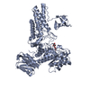
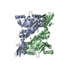
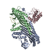
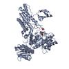
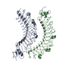
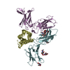
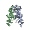
| Title | Crystal structure of the heterotrimer core of the S. cerevisiae AMPK homolog SNF1 | ||||||
 Components Components |
| ||||||
 Keywords Keywords | Transferase/Protein binding / heterotrimer / ATP-binding / Carbohydrate metabolism / Kinase / Membrane / Nucleotide-binding / Nucleus / Phosphorylation / Serine/threonine-protein kinase / Transferase / Lipoprotein / Myristate / CBS domain / Transcription / Transcription regulation / Transferase-Protein binding COMPLEX | ||||||
| Function / homology |  Function and homology information Function and homology informationfungal-type cell wall assembly / positive regulation of pseudohyphal growth / AMPK inhibits chREBP transcriptional activation activity / Energy dependent regulation of mTOR by LKB1-AMPK / regulation of cellular response to glucose starvation / single-species surface biofilm formation / positive regulation of filamentous growth of a population of unicellular organisms in response to starvation / regulation of invasive growth in response to glucose limitation / cellular bud neck septin ring / Carnitine shuttle ...fungal-type cell wall assembly / positive regulation of pseudohyphal growth / AMPK inhibits chREBP transcriptional activation activity / Energy dependent regulation of mTOR by LKB1-AMPK / regulation of cellular response to glucose starvation / single-species surface biofilm formation / positive regulation of filamentous growth of a population of unicellular organisms in response to starvation / regulation of invasive growth in response to glucose limitation / cellular bud neck septin ring / Carnitine shuttle / negative regulation of inositol phosphate biosynthetic process / invasive growth in response to glucose limitation / Macroautophagy / AMP-activated protein kinase activity / regulation of carbon utilization / peroxisome organization / filamentous growth / nucleotide-activated protein kinase complex / protein kinase regulator activity / regulation of glycolytic process / vacuolar membrane / cellular response to stress / AMP binding / nuclear envelope lumen / establishment of mitotic spindle orientation / positive regulation of macroautophagy / response to unfolded protein / regulation of protein-containing complex assembly / enzyme-substrate adaptor activity / cellular response to glucose starvation / guanyl-nucleotide exchange factor activity / positive regulation of gluconeogenesis / response to endoplasmic reticulum stress / molecular function activator activity / protein serine/threonine kinase activator activity / autophagy / nuclear membrane / protein kinase activity / non-specific serine/threonine protein kinase / negative regulation of translation / protein serine kinase activity / protein serine/threonine kinase activity / regulation of transcription by RNA polymerase II / protein kinase binding / signal transduction / mitochondrion / ATP binding / identical protein binding / nucleus / plasma membrane / cytoplasm / cytosol Similarity search - Function | ||||||
| Biological species |  | ||||||
| Method |  X-RAY DIFFRACTION / SYNCHROTRON / MOLECULAR REPLACEMENT / Resolution: 2.6 Å X-RAY DIFFRACTION / SYNCHROTRON / MOLECULAR REPLACEMENT / Resolution: 2.6 Å | ||||||
 Authors Authors | Amodeo, G.A. / Rudolph, M.J. / Tong, L. | ||||||
 Citation Citation | Journal: Nature / Year: 2007 Title: Crystal structure of the heterotrimer core of Saccharomyces cerevisiae AMPK homologue SNF1. Authors: Amodeo, G.A. / Rudolph, M.J. / Tong, L. | ||||||
| History |
|
- Structure visualization
| Structure viewer | Molecule: MolmilJmol/JSmol |
|---|
- Downloads & links
-Download
| PDBx/mmCIF format | 2qlv.cif.gz | 248.6 KB | Display | PDBx/mmCIF format |
|---|---|---|---|---|
| PDB format | pdb2qlv.ent.gz | 197.4 KB | Display | PDB format |
| PDBx/mmJSON format | 2qlv.json.gz | Tree view | PDBx/mmJSON format | |
| Others |  Other downloads Other downloads |
-Validation report
| Arichive directory | https://data.pdbj.org/pub/pdb/validation_reports/ql/2qlvftp://data.pdbj.org/pub/pdb/validation_reports/ql/2qlv | HTTPS FTP |
|---|
-Related structure data
| Similar structure data |
|---|


-Links
 PDBj
PDBj

- Assembly
| Deposited unit | 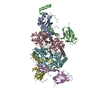
| ||||||||
|---|---|---|---|---|---|---|---|---|---|
| 1 | 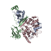
| ||||||||
| 2 | 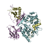
| ||||||||
| Unit cell |
| ||||||||
| Details | The biological assembly is that of a trimer consisting of Snf1, Sip2, and Snf4. |
-Components
| #1: Protein | Mass: 19596.268 Da / Num. of mol.: 2 Source method: isolated from a genetically manipulated source Source: (gene. exp.) Gene: SNF1, CAT1, CCR1, GLC2, PAS14 / Plasmid: pET 28a / Production host:  References: UniProt: P06782, non-specific serine/threonine protein kinase #2: Protein | Mass: 28956.588 Da / Num. of mol.: 2 Source method: isolated from a genetically manipulated source Source: (gene. exp.) Gene: SIP2, SPM2 / Plasmid: pET 28a / Production host: #3: Protein | Mass: 35625.066 Da / Num. of mol.: 2 Source method: isolated from a genetically manipulated source Source: (gene. exp.) Gene: SNF4, CAT3 / Plasmid: pET 28a / Production host: #4: Water | ChemComp-HOH / |  Mass: 18.015 Da / Num. of mol.: 111 / Source method: isolated from a natural source / Formula: H2O Mass: 18.015 Da / Num. of mol.: 111 / Source method: isolated from a natural source / Formula: H2O |
|---|
-Experimental details
-Experiment
| Experiment | Method: X-RAY DIFFRACTION / Number of used crystals: 1 |
|---|
- Sample preparation
| Crystal | Density Matthews: 2.33 Å3/Da / Density % sol: 47.24 % |
|---|---|
| Crystal grow | Temperature: 294 K / Method: vapor diffusion, hanging drop / pH: 7 Details: 250 mM ammonium citrate, 15% PEG3350, 1mM AMP, pH 7.0, VAPOR DIFFUSION, HANGING DROP, temperature 294K |
-Data collection
| Diffraction | Mean temperature: 100 K | |||||||||||||||||||||||||||||||||||||||||||||||||||||||||||||||||||||||||||||
|---|---|---|---|---|---|---|---|---|---|---|---|---|---|---|---|---|---|---|---|---|---|---|---|---|---|---|---|---|---|---|---|---|---|---|---|---|---|---|---|---|---|---|---|---|---|---|---|---|---|---|---|---|---|---|---|---|---|---|---|---|---|---|---|---|---|---|---|---|---|---|---|---|---|---|---|---|---|---|
| Diffraction source | Source: SYNCHROTRON / Site: NSLS  / Beamline: X29A / Wavelength: 1.0809 Å / Beamline: X29A / Wavelength: 1.0809 Å | |||||||||||||||||||||||||||||||||||||||||||||||||||||||||||||||||||||||||||||
| Detector | Type: ADSC QUANTUM 315 / Detector: CCD / Date: Apr 21, 2007 | |||||||||||||||||||||||||||||||||||||||||||||||||||||||||||||||||||||||||||||
| Radiation | Monochromator: Cryogenically cooled double crystal monochrometer with horizontal focusing sagittal bend second mono crystal with 4:1 magnification ratio and vertically focusing mirror Protocol: SINGLE WAVELENGTH / Monochromatic (M) / Laue (L): M / Scattering type: x-ray | |||||||||||||||||||||||||||||||||||||||||||||||||||||||||||||||||||||||||||||
| Radiation wavelength | Wavelength: 1.0809 Å / Relative weight: 1 | |||||||||||||||||||||||||||||||||||||||||||||||||||||||||||||||||||||||||||||
| Reflection | Redundancy: 2.9 % / Av σ(I) over netI: 17.4 / Number: 130084 / Rmerge(I) obs: 0.071 / Χ2: 1.02 / D res high: 2.6 Å / D res low: 30 Å / Num. obs: 44415 / % possible obs: 93.3 | |||||||||||||||||||||||||||||||||||||||||||||||||||||||||||||||||||||||||||||
| Diffraction reflection shell |
| |||||||||||||||||||||||||||||||||||||||||||||||||||||||||||||||||||||||||||||
| Reflection | Resolution: 2.6→30 Å / Num. obs: 44415 / % possible obs: 93.3 % / Redundancy: 2.9 % / Rmerge(I) obs: 0.071 / Χ2: 1.018 / Net I/σ(I): 17.4 | |||||||||||||||||||||||||||||||||||||||||||||||||||||||||||||||||||||||||||||
| Reflection shell | Resolution: 2.6→2.69 Å / Redundancy: 2.1 % / Rmerge(I) obs: 0.385 / Num. unique all: 3361 / Χ2: 1.075 / % possible all: 71.3 |
- Processing
| Software |
| ||||||||||||||||||||||||||||||||||||
|---|---|---|---|---|---|---|---|---|---|---|---|---|---|---|---|---|---|---|---|---|---|---|---|---|---|---|---|---|---|---|---|---|---|---|---|---|---|
| Refinement | Method to determine structure: MOLECULAR REPLACEMENT / Resolution: 2.6→29.54 Å / Rfactor Rfree error: 0.005 / FOM work R set: 0.727 / Data cutoff high absF: 501624.375 / Data cutoff low absF: 0 / Isotropic thermal model: RESTRAINED / Cross valid method: THROUGHOUT / σ(F): 0.3
| ||||||||||||||||||||||||||||||||||||
| Solvent computation | Solvent model: FLAT MODEL / Bsol: 45.919 Å2 / ksol: 0.311 e/Å3 | ||||||||||||||||||||||||||||||||||||
| Displacement parameters | Biso mean: 66.3 Å2
| ||||||||||||||||||||||||||||||||||||
| Refine analyze |
| ||||||||||||||||||||||||||||||||||||
| Refinement step | Cycle: LAST / Resolution: 2.6→29.54 Å
| ||||||||||||||||||||||||||||||||||||
| Refine LS restraints |
| ||||||||||||||||||||||||||||||||||||
| LS refinement shell | Resolution: 2.6→2.69 Å / Rfactor Rfree error: 0.027 / Total num. of bins used: 10
| ||||||||||||||||||||||||||||||||||||
| Xplor file |
|
