 Movie
Movie Controller

[English] 日本語
 Yorodumi
- PDB-1n9w: Crystal structure of the non-discriminating and archaeal-type asp... -
+ Open data

- Basic information
| Entry | Database: PDB / ID: 1n9w | ||||||
|---|---|---|---|---|---|---|---|
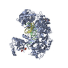
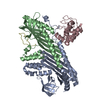
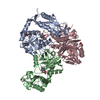
| Title | Crystal structure of the non-discriminating and archaeal-type aspartyl-tRNA synthetase from Thermus thermophilus | ||||||
 Components Components | aspartyl-tRNA synthetase 2 | ||||||
 Keywords Keywords | BIOSYNTHETIC PROTEIN | ||||||
| Function / homology |  Function and homology information Function and homology informationaspartate-tRNAAsn ligase / aspartate-tRNA(Asn) ligase activity / aspartate-tRNA ligase activity / aspartyl-tRNA aminoacylation / aminoacyl-tRNA synthetase multienzyme complex / RNA binding / ATP binding / cytosol Similarity search - Function | ||||||
| Biological species |   Thermus thermophilus (bacteria) Thermus thermophilus (bacteria) | ||||||
| Method |  X-RAY DIFFRACTION / SYNCHROTRON / MOLECULAR REPLACEMENT / Resolution: 2.3 Å X-RAY DIFFRACTION / SYNCHROTRON / MOLECULAR REPLACEMENT / Resolution: 2.3 Å | ||||||
 Authors Authors | Charron, C. / Roy, H. / Blaise, M. / Giege, R. / Kern, D. | ||||||
 Citation Citation | Journal: EMBO J. / Year: 2003 Title: Non-discriminating and discriminating aspartyl-tRNA synthetases differ in the anticodon-binding domain Authors: Charron, C. / Roy, H. / Blaise, M. / Giege, R. / Kern, D. #1: Journal: Acta Crystallogr.,Sect.D / Year: 2001Title: Crystallization and preliminary X-ray diffraction data of the second and archaebacterial-type aspartyl-tRNA synthetase from Thermus thermophilus Authors: Charron, C. / Roy, H. / Lorber, L. / Kern, D. / Giege, R. | ||||||
| History |
|
- Structure visualization
| Structure viewer | Molecule: MolmilJmol/JSmol |
|---|
- Downloads & links
-Download
| PDBx/mmCIF format | 1n9w.cif.gz | 153.9 KB | Display | PDBx/mmCIF format |
|---|---|---|---|---|
| PDB format | pdb1n9w.ent.gz | 122.9 KB | Display | PDB format |
| PDBx/mmJSON format | 1n9w.json.gz | Tree view | PDBx/mmJSON format | |
| Others |  Other downloads Other downloads |
-Validation report
| Arichive directory | https://data.pdbj.org/pub/pdb/validation_reports/n9/1n9wftp://data.pdbj.org/pub/pdb/validation_reports/n9/1n9w | HTTPS FTP |
|---|
-Related structure data
| Similar structure data |
|---|


-Links
 PDBj
PDBj

- Assembly
| Deposited unit | 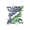
| ||||||||
|---|---|---|---|---|---|---|---|---|---|
| 1 |
| ||||||||
| Unit cell |
| ||||||||
| Details | The biological assembly is a dimer in the asymmetric unit |
-Components
| #1: Protein | Mass: 48397.262 Da / Num. of mol.: 2 Source method: isolated from a genetically manipulated source Source: (gene. exp.) Thermus thermophilus (bacteria) / Production host: #2: Water | ChemComp-HOH / |  Mass: 18.015 Da / Num. of mol.: 159 / Source method: isolated from a natural source / Formula: H2O Mass: 18.015 Da / Num. of mol.: 159 / Source method: isolated from a natural source / Formula: H2O |
|---|
-Experimental details
-Experiment
| Experiment | Method: X-RAY DIFFRACTION / Number of used crystals: 1 |
|---|
- Sample preparation
| Crystal | Density Matthews: 3.03 Å3/Da / Density % sol: 59.45 % | ||||||||||||||||||||||||||||
|---|---|---|---|---|---|---|---|---|---|---|---|---|---|---|---|---|---|---|---|---|---|---|---|---|---|---|---|---|---|
| Crystal grow | Temperature: 293 K / Method: vapor diffusion, hanging drop / pH: 9.5 Details: PEG 8000 and NaCl, pH 9.5, VAPOR DIFFUSION, HANGING DROP, temperature 293K | ||||||||||||||||||||||||||||
| Crystal grow | *PLUS Method: vapor diffusion | ||||||||||||||||||||||||||||
| Components of the solutions | *PLUS
|
-Data collection
| Diffraction | Mean temperature: 100 K |
|---|---|
| Diffraction source | Source: SYNCHROTRON / Site: ESRF  / Beamline: ID14-4 / Wavelength: 0.933 Å / Beamline: ID14-4 / Wavelength: 0.933 Å |
| Detector | Type: ADSC QUANTUM 4 / Detector: CCD / Date: Oct 10, 2001 |
| Radiation | Protocol: SINGLE WAVELENGTH / Monochromatic (M) / Laue (L): M / Scattering type: x-ray |
| Radiation wavelength | Wavelength: 0.933 Å / Relative weight: 1 |
| Reflection | Resolution: 2.3→30 Å / Num. all: 53223 / Num. obs: 52274 / % possible obs: 98.2 % / Observed criterion σ(F): 2 / Observed criterion σ(I): 0 / Redundancy: 4.2 % / Rsym value: 0.045 / Net I/σ(I): 11.5 |
| Reflection shell | Resolution: 2.3→2.36 Å / Redundancy: 4.4 % / Mean I/σ(I) obs: 3.1 / Rsym value: 0.221 / % possible all: 99.9 |
| Reflection | *PLUS Lowest resolution: 30 Å / % possible obs: 99.8 % / Rmerge(I) obs: 0.045 |
| Reflection shell | *PLUS Highest resolution: 2.3 Å / % possible obs: 99.9 % / Rmerge(I) obs: 0.221 |
- Processing
| Software |
| ||||||||||||||||||||
|---|---|---|---|---|---|---|---|---|---|---|---|---|---|---|---|---|---|---|---|---|---|
| Refinement | Method to determine structure: MOLECULAR REPLACEMENT / Resolution: 2.3→30 Å / σ(F): 2 / Stereochemistry target values: Engh & Huber
| ||||||||||||||||||||
| Refinement step | Cycle: LAST / Resolution: 2.3→30 Å
| ||||||||||||||||||||
| Refine LS restraints |
| ||||||||||||||||||||
| LS refinement shell | Resolution: 2.3→2.36 Å
| ||||||||||||||||||||
| Refinement | *PLUS Lowest resolution: 30 Å / Num. reflection obs: 45048 / % reflection Rfree: 7 % | ||||||||||||||||||||
| Solvent computation | *PLUS | ||||||||||||||||||||
| Displacement parameters | *PLUS | ||||||||||||||||||||
| Refine LS restraints | *PLUS Type: c_angle_deg / Dev ideal: 1.2 | ||||||||||||||||||||
| LS refinement shell | *PLUS Highest resolution: 2.3 Å |
