Entry Database : PDB / ID : 5cm0Title Crystal structure of branched-chain aminotransferase from thermophilic archaea Geoglobus acetivorans Branched-chain transaminase Keywords / / / / / Function / homology Function Domain/homology Component
/ / / / / / / / / / / / / / / / / / / / / / / / / / / / / / Biological species Geoglobus acetivorans (archaea)Method / / / / Resolution : 1.9 Å Authors Boyko, K.M. / Nikolaeva, A.Y. / Stekhanova, T.N. / Mardanov, A.V. / Rakitin, A.L. / Ravin, N.V. / Popov, V.O. Funding support Organization Grant number Country Russian Scientific Fund ERA-IB ("Thermogene") 7th EU Framework Programme
Journal : Front Bioeng Biotechnol / Year : 2019Title : Thermostable Branched-Chain Amino Acid Transaminases From the Archaea Geoglobus acetivorans and Archaeoglobus fulgidus : Biochemical and Structural Characterization.Authors: Isupov, M.N. / Boyko, K.M. / Sutter, J.M. / James, P. / Sayer, C. / Schmidt, M. / Schonheit, P. / Nikolaeva, A.Y. / Stekhanova, T.N. / Mardanov, A.V. / Ravin, N.V. / Bezsudnova, E.Y. / ... Authors : Isupov, M.N. / Boyko, K.M. / Sutter, J.M. / James, P. / Sayer, C. / Schmidt, M. / Schonheit, P. / Nikolaeva, A.Y. / Stekhanova, T.N. / Mardanov, A.V. / Ravin, N.V. / Bezsudnova, E.Y. / Popov, V.O. / Littlechild, J.A. History Deposition Jul 16, 2015 Deposition site / Processing site Revision 1.0 Sep 14, 2016 Provider / Type Revision 1.1 Mar 15, 2017 Group Revision 1.2 Mar 31, 2021 Group / Derived calculations / Structure summaryCategory citation / citation_author ... citation / citation_author / entity / pdbx_struct_special_symmetry Item _citation.journal_abbrev / _citation.journal_id_CSD ... _citation.journal_abbrev / _citation.journal_id_CSD / _citation.journal_id_ISSN / _citation.journal_volume / _citation.page_first / _citation.page_last / _citation.pdbx_database_id_DOI / _citation.pdbx_database_id_PubMed / _citation.title / _citation.year / _entity.pdbx_ec Revision 1.3 Jan 10, 2024 Group / Database references / Refinement descriptionCategory chem_comp_atom / chem_comp_bond ... chem_comp_atom / chem_comp_bond / database_2 / pdbx_initial_refinement_model Item / _database_2.pdbx_database_accession
Show all Show less
 Movie
Movie Controller

 Yorodumi
 Open data
 Basic information
 Components
 Keywords
 Function and homology information
 Geoglobus acetivorans (archaea)
 X-RAY DIFFRACTION /
  Authors
 Russian Federation, 2items
  Citation
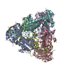
 Structure visualization
 Downloads & links
 Other downloads


 PDBj
PDBj
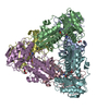
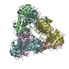
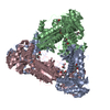
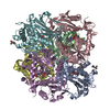
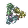
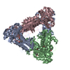
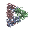
 Assembly
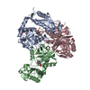


 Mass: 247.142 Da / Num. of mol.: 3 / Source method: isolated from a natural source / Formula: C8H10NO6P
 Mass: 92.094 Da / Num. of mol.: 2 / Source method: isolated from a natural source / Formula: C3H8O3
 Mass: 18.015 Da / Num. of mol.: 626 / Source method: isolated from a natural source / Formula: H2O
 Sample preparation
 / Beamline: BL41XU / Wavelength: 1 Å
 Processing