 Movie
Movie Controller

[English] 日本語
 Yorodumi
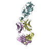
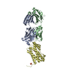
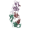
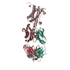
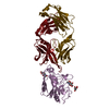
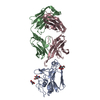
- PDB-7cm4: Crystal Structure of COVID-19 virus spike receptor-binding domain... -
+ Open data

- Basic information
| Entry | Database: PDB / ID: 7cm4 | ||||||
|---|---|---|---|---|---|---|---|
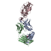
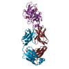
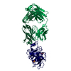
| Title | Crystal Structure of COVID-19 virus spike receptor-binding domain complexed with a neutralizing antibody CT-P59 | ||||||
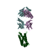
 Components Components |
| ||||||
 Keywords Keywords | VIRAL PROTEIN / SARS-Cov-2 / spike / receptor binding domain / RBD / IgG / Fab | ||||||
| Function / homology |  Function and homology information Function and homology informationsymbiont-mediated disruption of host tissue / Maturation of spike protein / Translation of Structural Proteins / Virion Assembly and Release / host cell surface / host extracellular region / symbiont-mediated-mediated suppression of host tetherin activity / Induction of Cell-Cell Fusion / structural constituent of virion / positive regulation of viral entry into host cell ...symbiont-mediated disruption of host tissue / Maturation of spike protein / Translation of Structural Proteins / Virion Assembly and Release / host cell surface / host extracellular region / symbiont-mediated-mediated suppression of host tetherin activity / Induction of Cell-Cell Fusion / structural constituent of virion / positive regulation of viral entry into host cell / membrane fusion / host cell endoplasmic reticulum-Golgi intermediate compartment membrane / Attachment and Entry / entry receptor-mediated virion attachment to host cell / receptor-mediated virion attachment to host cell / host cell surface receptor binding / symbiont-mediated suppression of host innate immune response / endocytosis involved in viral entry into host cell / receptor ligand activity / fusion of virus membrane with host plasma membrane / fusion of virus membrane with host endosome membrane / viral envelope / symbiont entry into host cell / virion attachment to host cell / host cell plasma membrane / SARS-CoV-2 activates/modulates innate and adaptive immune responses / virion membrane / membrane / identical protein binding / plasma membrane Similarity search - Function | ||||||
| Biological species |   Severe acute respiratory syndrome coronavirus 2 Severe acute respiratory syndrome coronavirus 2 Homo sapiens (human) Homo sapiens (human) | ||||||
| Method |  X-RAY DIFFRACTION / SYNCHROTRON / MOLECULAR REPLACEMENT / Resolution: 2.71 Å X-RAY DIFFRACTION / SYNCHROTRON / MOLECULAR REPLACEMENT / Resolution: 2.71 Å | ||||||
 Authors Authors | Kim, Y.G. / Jeong, J.H. / Bae, J.S. / Lee, J. | ||||||
 Citation Citation | Journal: Nat Commun / Year: 2021 Title: A therapeutic neutralizing antibody targeting receptor binding domain of SARS-CoV-2 spike protein. Authors: Kim, C. / Ryu, D.K. / Lee, J. / Kim, Y.I. / Seo, J.M. / Kim, Y.G. / Jeong, J.H. / Kim, M. / Kim, J.I. / Kim, P. / Bae, J.S. / Shim, E.Y. / Lee, M.S. / Kim, M.S. / Noh, H. / Park, G.S. / ...Authors: Kim, C. / Ryu, D.K. / Lee, J. / Kim, Y.I. / Seo, J.M. / Kim, Y.G. / Jeong, J.H. / Kim, M. / Kim, J.I. / Kim, P. / Bae, J.S. / Shim, E.Y. / Lee, M.S. / Kim, M.S. / Noh, H. / Park, G.S. / Park, J.S. / Son, D. / An, Y. / Lee, J.N. / Kwon, K.S. / Lee, J.Y. / Lee, H. / Yang, J.S. / Kim, K.C. / Kim, S.S. / Woo, H.M. / Kim, J.W. / Park, M.S. / Yu, K.M. / Kim, S.M. / Kim, E.H. / Park, S.J. / Jeong, S.T. / Yu, C.H. / Song, Y. / Gu, S.H. / Oh, H. / Koo, B.S. / Hong, J.J. / Ryu, C.M. / Park, W.B. / Oh, M.D. / Choi, Y.K. / Lee, S.Y. | ||||||
| History |
|
- Structure visualization
| Structure viewer | Molecule: MolmilJmol/JSmol |
|---|
- Downloads & links
-Download
| PDBx/mmCIF format | 7cm4.cif.gz | 167.9 KB | Display | PDBx/mmCIF format |
|---|---|---|---|---|
| PDB format | pdb7cm4.ent.gz | 108.4 KB | Display | PDB format |
| PDBx/mmJSON format | 7cm4.json.gz | Tree view | PDBx/mmJSON format | |
| Others |  Other downloads Other downloads |
-Validation report
| Arichive directory | https://data.pdbj.org/pub/pdb/validation_reports/cm/7cm4ftp://data.pdbj.org/pub/pdb/validation_reports/cm/7cm4 | HTTPS FTP |
|---|
-Related structure data
| Related structure data | 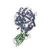 6lzgS S: Starting model for refinement |
|---|---|
| Similar structure data |


-Links
 PDBj
PDBj


- Assembly
| Deposited unit | 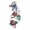
| ||||||||||||
|---|---|---|---|---|---|---|---|---|---|---|---|---|---|
| 1 |
| ||||||||||||
| Unit cell |
|
-Components
-Antibody , 2 types, 2 molecules HL
| #2: Antibody | Mass: 50547.078 Da / Num. of mol.: 1 Source method: isolated from a genetically manipulated source Source: (gene. exp.) Homo sapiens (human) / Production host:   Cricetulus griseus (Chinese hamster) Cricetulus griseus (Chinese hamster) |
|---|---|
| #3: Antibody | Mass: 22655.049 Da / Num. of mol.: 1 Source method: isolated from a genetically manipulated source Source: (gene. exp.) Homo sapiens (human) / Production host: Cricetulus griseus (Chinese hamster) |
-Protein / Sugars , 2 types, 2 molecules A
| #1: Protein | Mass: 25634.781 Da / Num. of mol.: 1 Source method: isolated from a genetically manipulated source Source: (gene. exp.) Severe acute respiratory syndrome coronavirus 2Gene: S, 2 / Production host:   Spodoptera frugiperda (fall armyworm) / References: UniProt: P0DTC2 Spodoptera frugiperda (fall armyworm) / References: UniProt: P0DTC2 |
|---|---|
| #4: Polysaccharide | alpha-D-mannopyranose-(1-6)-alpha-D-mannopyranose-(1-4)-2-acetamido-2-deoxy-beta-D-glucopyranose-(1- ...alpha-D-mannopyranose-(1-6)-alpha-D-mannopyranose-(1-4)-2-acetamido-2-deoxy-beta-D-glucopyranose-(1-4)-[alpha-L-fucopyranose-(1-6)]2-acetamido-2-deoxy-beta-D-glucopyranose Source method: isolated from a genetically manipulated source |
-Non-polymers , 3 types, 69 molecules 


| #5: Chemical |  Mass: 58.693 Da / Num. of mol.: 2 / Source method: obtained synthetically / Formula: Ni Mass: 58.693 Da / Num. of mol.: 2 / Source method: obtained synthetically / Formula: Ni#6: Chemical |  Mass: 62.068 Da / Num. of mol.: 2 / Source method: obtained synthetically / Formula: C2H6O2 Mass: 62.068 Da / Num. of mol.: 2 / Source method: obtained synthetically / Formula: C2H6O2#7: Water | ChemComp-HOH / | Mass: 18.015 Da / Num. of mol.: 65 / Source method: isolated from a natural source / Formula: H2O |
|---|
-Details
| Has ligand of interest | N |
|---|---|
| Has protein modification | Y |
-Experimental details
-Experiment
| Experiment | Method: X-RAY DIFFRACTION / Number of used crystals: 1 |
|---|
- Sample preparation
| Crystal | Density Matthews: 2.35 Å3/Da / Density % sol: 47.63 % |
|---|---|
| Crystal grow | Temperature: 293.15 K / Method: vapor diffusion, sitting drop / pH: 8 Details: 100 mM Tris-Cl pH 8.0 16% (w/v) Polyethylene glycol monomethyl ether 2,000 10 mM Nickel(II) Chloride hexahydrate |
-Data collection
| Diffraction | Mean temperature: 100 K / Serial crystal experiment: N |
|---|---|
| Diffraction source | Source: SYNCHROTRON / Site: PAL/PLS  / Beamline: 5C (4A) / Wavelength: 0.9796 Å / Beamline: 5C (4A) / Wavelength: 0.9796 Å |
| Detector | Type: DECTRIS EIGER X 9M / Detector: PIXEL / Date: Jul 7, 2020 |
| Radiation | Monochromator: Double Crystal Monochromator / Protocol: SINGLE WAVELENGTH / Monochromatic (M) / Laue (L): M / Scattering type: x-ray |
| Radiation wavelength | Wavelength: 0.9796 Å / Relative weight: 1 |
| Reflection | Resolution: 2.71→29.76 Å / Num. obs: 25744 / % possible obs: 99.78 % / Redundancy: 13.6 % / Biso Wilson estimate: 58.06 Å2 / CC1/2: 0.997 / CC star: 0.999 / Rpim(I) all: 0.06856 / Rrim(I) all: 0.2543 / Net I/σ(I): 12.44 |
| Reflection shell | Resolution: 2.71→2.807 Å / Redundancy: 99.88 % / Mean I/σ(I) obs: 1.63 / Num. unique obs: 2541 / CC1/2: 0.606 / CC star: 0.869 / Rpim(I) all: 0.6129 / Rrim(I) all: 2.232 |
- Processing
| Software |
| |||||||||||||||||||||||||||||||||||||||||||||||||||||||||||||||||||||||||||||||||||||||||||||||||||||||||
|---|---|---|---|---|---|---|---|---|---|---|---|---|---|---|---|---|---|---|---|---|---|---|---|---|---|---|---|---|---|---|---|---|---|---|---|---|---|---|---|---|---|---|---|---|---|---|---|---|---|---|---|---|---|---|---|---|---|---|---|---|---|---|---|---|---|---|---|---|---|---|---|---|---|---|---|---|---|---|---|---|---|---|---|---|---|---|---|---|---|---|---|---|---|---|---|---|---|---|---|---|---|---|---|---|---|---|
| Refinement | Method to determine structure: MOLECULAR REPLACEMENT Starting model: 6LZG Resolution: 2.71→29.76 Å / SU ML: 0.3896 / Cross valid method: FREE R-VALUE / σ(F): 1.35 / Phase error: 26.4007 Stereochemistry target values: GeoStd + Monomer Library + CDL v1.2
| |||||||||||||||||||||||||||||||||||||||||||||||||||||||||||||||||||||||||||||||||||||||||||||||||||||||||
| Solvent computation | Shrinkage radii: 0.9 Å / VDW probe radii: 1.11 Å / Solvent model: FLAT BULK SOLVENT MODEL | |||||||||||||||||||||||||||||||||||||||||||||||||||||||||||||||||||||||||||||||||||||||||||||||||||||||||
| Displacement parameters | Biso mean: 65.14 Å2 | |||||||||||||||||||||||||||||||||||||||||||||||||||||||||||||||||||||||||||||||||||||||||||||||||||||||||
| Refinement step | Cycle: LAST / Resolution: 2.71→29.76 Å
| |||||||||||||||||||||||||||||||||||||||||||||||||||||||||||||||||||||||||||||||||||||||||||||||||||||||||
| Refine LS restraints |
| |||||||||||||||||||||||||||||||||||||||||||||||||||||||||||||||||||||||||||||||||||||||||||||||||||||||||
| LS refinement shell |
|
