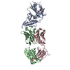
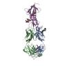
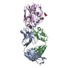
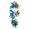
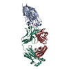
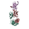
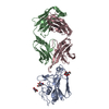
S: Voltage-sensor containing phosphatase A: fragment antibody heavy chain B: fragment antibody light chain C: fragment antibody heavy chain D: fragment antibody light chain T: Voltage-sensor containing phosphatase E: fragment antibody heavy chain F: fragment antibody light chain G: fragment antibody heavy chain H: fragment antibody light chain I: Voltage-sensor containing phosphatase J: Voltage-sensor containing phosphatase
Mass: 17750.729 Da / Num. of mol.: 4 Source method: isolated from a genetically manipulated source Source: (gene. exp.) Ciona intestinalis (vase tunicate) / Gene: Ci-VSP / Plasmid: pQE32 with sequence encoding for Ci-VSD / Production host: Escherichia coli (E. coli) / Strain (production host): XL10-Gold / References: UniProt: Q4W8A1, UniProt: F6XHE4*PLUS
#2: Antibody
fragmentantibodyheavychain
Mass: 23988.711 Da / Num. of mol.: 4 Source method: isolated from a genetically manipulated source Source: (gene. exp.) Homo sapiens (human) Plasmid: phagemid coding for both heavy and light chain of the fragment antibody Production host: Escherichia coli (E. coli) / Strain (production host): 55244
#3: Antibody
fragmentantibodylightchain
Mass: 22989.506 Da / Num. of mol.: 4 Source method: isolated from a genetically manipulated source Source: (gene. exp.) Homo sapiens (human) Plasmid: phagemid coding for both heavy and light chain of the fragment antibody Production host: Escherichia coli (E. coli) / Strain (production host): 55244
Has protein modification
Y
-
Experimental details
-
Experiment
Experiment
Method: X-RAY DIFFRACTION / Number of used crystals: 40
-
Sample preparation
Crystal
Density Matthews: 5.1 Å3/Da / Density % sol: 75.89 %
Crystal grow
Temperature: 293 K / Method: vapor diffusion, hanging drop / pH: 5.5 Details: 0.1 M Na Citrate, 0.1 M Li2SO4, 20% PEG 1000, 10% glycerol, pH 5.5, VAPOR DIFFUSION, HANGING DROP, temperature 293K
Method to determine structure: MOLECULAR REPLACEMENT / Resolution: 3.58→50 Å / Cor.coef. Fo:Fc: 0.902 / Cor.coef. Fo:Fc free: 0.871 / SU B: 39.019 / SU ML: 0.555 / Cross valid method: THROUGHOUT / ESU R Free: 0.627 / Stereochemistry target values: MAXIMUM LIKELIHOOD / Details: HYDROGENS HAVE BEEN USED IF PRESENT IN THE INPUT
Rfactor
Num. reflection
% reflection
Selection details
Rfree
0.29225
2803
5 %
RANDOM
Rwork
0.24794
-
-
-
obs
0.25022
52942
92.57 %
-
Solvent computation
Ion probe radii: 0.8 Å / Shrinkage radii: 0.8 Å / VDW probe radii: 1.2 Å / Solvent model: MASK
 Movie
Movie Controller

 Yorodumi
 Open data
 Basic information
 Components
 Keywords
 Function and homology information
 Homo sapiens (human)
 X-RAY DIFFRACTION /
  Authors
 Citation
 Structure visualization
 Downloads & links
 Other downloads
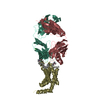


 PDBj
PDBj


 Assembly


 Sample preparation
 Processing