 Movie
Movie Controller

[English] 日本語
 Yorodumi
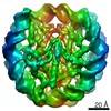
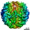
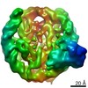
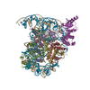
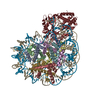
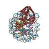
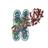
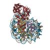
- PDB-7bxt: The cryo-EM structure of CENP-A nucleosome in complex with CENP-C... -
+ Open data

- Basic information
| Entry | Database: PDB / ID: 7bxt | ||||||||||||
|---|---|---|---|---|---|---|---|---|---|---|---|---|---|
| Title | The cryo-EM structure of CENP-A nucleosome in complex with CENP-C peptide and CENP-N N-terminal domain | ||||||||||||
 Components Components |
| ||||||||||||
 Keywords Keywords | CELL CYCLE/DNA / CENP-A nucleosome / CENP-C / CENP-N / complex / kinetochore / NUCLEAR PROTEIN / CELL CYCLE-DNA complex | ||||||||||||
| Function / homology |  Function and homology information Function and homology informationkinetochore => GO:0000776 / Interleukin-7 signaling / Amplification of signal from unattached kinetochores via a MAD2 inhibitory signal / Chromatin modifying enzymes / Formation of the beta-catenin:TCF transactivating complex / : / Transcriptional regulation by small RNAs / : / RHO GTPases Activate Formins / PRC2 methylates histones and DNA ...kinetochore => GO:0000776 / Interleukin-7 signaling / Amplification of signal from unattached kinetochores via a MAD2 inhibitory signal / Chromatin modifying enzymes / Formation of the beta-catenin:TCF transactivating complex / : / Transcriptional regulation by small RNAs / : / RHO GTPases Activate Formins / PRC2 methylates histones and DNA / Oxidative Stress Induced Senescence / HATs acetylate histones / Assembly of the ORC complex at the origin of replication / RNA Polymerase I Promoter Opening / RNA Polymerase I Promoter Escape / RUNX1 regulates genes involved in megakaryocyte differentiation and platelet function / Estrogen-dependent gene expression / Negative Regulation of CDH1 Gene Transcription / MLL4 and MLL3 complexes regulate expression of PPARG target genes in adipogenesis and hepatic steatosis / Regulation of endogenous retroelements by KRAB-ZFP proteins / Regulation of endogenous retroelements by the Human Silencing Hub (HUSH) complex / Resolution of Sister Chromatid Cohesion / B-WICH complex positively regulates rRNA expression / EML4 and NUDC in mitotic spindle formation / Deposition of new CENPA-containing nucleosomes at the centromere / Separation of Sister Chromatids / Factors involved in megakaryocyte development and platelet production / spindle attachment to meiosis I kinetochore / centromeric DNA binding / CENP-A containing chromatin assembly / protein localization to chromosome, centromeric region / kinetochore assembly / attachment of mitotic spindle microtubules to kinetochore / condensed chromosome, centromeric region / establishment of mitotic spindle orientation / detection of maltose stimulus / maltose transport complex / mitotic cytokinesis / carbohydrate transport / chromosome, centromeric region / carbohydrate transmembrane transporter activity / maltose binding / maltose transport / maltodextrin transmembrane transport / pericentric heterochromatin / negative regulation of megakaryocyte differentiation / ATP-binding cassette (ABC) transporter complex, substrate-binding subunit-containing / protein localization to CENP-A containing chromatin / Replacement of protamines by nucleosomes in the male pronucleus / CENP-A containing nucleosome / Packaging Of Telomere Ends / Recognition and association of DNA glycosylase with site containing an affected purine / Cleavage of the damaged purine / Deposition of new CENPA-containing nucleosomes at the centromere / telomere organization / ATP-binding cassette (ABC) transporter complex / Recognition and association of DNA glycosylase with site containing an affected pyrimidine / Cleavage of the damaged pyrimidine / RNA Polymerase I Promoter Opening / Inhibition of DNA recombination at telomere / Assembly of the ORC complex at the origin of replication / Meiotic synapsis / SUMOylation of chromatin organization proteins / Regulation of endogenous retroelements by the Human Silencing Hub (HUSH) complex / DNA methylation / Condensation of Prophase Chromosomes / Chromatin modifications during the maternal to zygotic transition (MZT) / SIRT1 negatively regulates rRNA expression / HCMV Late Events / ERCC6 (CSB) and EHMT2 (G9a) positively regulate rRNA expression / PRC2 methylates histones and DNA / Regulation of endogenous retroelements by KRAB-ZFP proteins / cell chemotaxis / Defective pyroptosis / innate immune response in mucosa / HDACs deacetylate histones / Regulation of endogenous retroelements by Piwi-interacting RNAs (piRNAs) / chromosome segregation / RNA Polymerase I Promoter Escape / Nonhomologous End-Joining (NHEJ) / Transcriptional regulation by small RNAs / HDMs demethylate histones / Formation of the beta-catenin:TCF transactivating complex / Activated PKN1 stimulates transcription of AR (androgen receptor) regulated genes KLK2 and KLK3 / RUNX1 regulates genes involved in megakaryocyte differentiation and platelet function / NoRC negatively regulates rRNA expression / Negative Regulation of CDH1 Gene Transcription / G2/M DNA damage checkpoint / kinetochore / PKMTs methylate histone lysines / B-WICH complex positively regulates rRNA expression / DNA Damage/Telomere Stress Induced Senescence / Meiotic recombination / Pre-NOTCH Transcription and Translation / Activation of anterior HOX genes in hindbrain development during early embryogenesis / Transcriptional regulation of granulopoiesis / RMTs methylate histone arginines / Metalloprotease DUBs / HCMV Early Events / structural constituent of chromatin Similarity search - Function | ||||||||||||
| Biological species |   Homo sapiens (human) Homo sapiens (human)unidentified cloning vector (others) synthetic construct (others) | ||||||||||||
| Method | ELECTRON MICROSCOPY / single particle reconstruction / cryo EM / Resolution: 4.2 Å | ||||||||||||
 Authors Authors | Ariyoshi, M. / Makino, F. / Fukagawa, T. | ||||||||||||
| Funding support |  Japan, 3items Japan, 3items
| ||||||||||||
 Citation Citation | Journal: EMBO J / Year: 2021 Title: Cryo-EM structure of the CENP-A nucleosome in complex with phosphorylated CENP-C. Authors: Mariko Ariyoshi / Fumiaki Makino / Reito Watanabe / Reiko Nakagawa / Takayuki Kato / Keiichi Namba / Yasuhiro Arimura / Risa Fujita / Hitoshi Kurumizaka / Ei-Ichi Okumura / Masatoshi Hara / Tatsuo Fukagawa / Abstract: The CENP-A nucleosome is a key structure for kinetochore assembly. Once the CENP-A nucleosome is established in the centromere, additional proteins recognize the CENP-A nucleosome to form a ...The CENP-A nucleosome is a key structure for kinetochore assembly. Once the CENP-A nucleosome is established in the centromere, additional proteins recognize the CENP-A nucleosome to form a kinetochore. CENP-C and CENP-N are CENP-A binding proteins. We previously demonstrated that vertebrate CENP-C binding to the CENP-A nucleosome is regulated by CDK1-mediated CENP-C phosphorylation. However, it is still unknown how the phosphorylation of CENP-C regulates its binding to CENP-A. It is also not completely understood how and whether CENP-C and CENP-N act together on the CENP-A nucleosome. Here, using cryo-electron microscopy (cryo-EM) in combination with biochemical approaches, we reveal a stable CENP-A nucleosome-binding mode of CENP-C through unique regions. The chicken CENP-C structure bound to the CENP-A nucleosome is stabilized by an intramolecular link through the phosphorylated CENP-C residue. The stable CENP-A-CENP-C complex excludes CENP-N from the CENP-A nucleosome. These findings provide mechanistic insights into the dynamic kinetochore assembly regulated by CDK1-mediated CENP-C phosphorylation. | ||||||||||||
| History |
|
- Structure visualization
| Movie |
Movie viewer |
|---|---|
| Structure viewer | Molecule: MolmilJmol/JSmol |
- Downloads & links
-Download
| PDBx/mmCIF format | 7bxt.cif.gz | 383 KB | Display | PDBx/mmCIF format |
|---|---|---|---|---|
| PDB format | pdb7bxt.ent.gz | 284.1 KB | Display | PDB format |
| PDBx/mmJSON format | 7bxt.json.gz | Tree view | PDBx/mmJSON format | |
| Others |  Other downloads Other downloads |
-Validation report
| Arichive directory | https://data.pdbj.org/pub/pdb/validation_reports/bx/7bxtftp://data.pdbj.org/pub/pdb/validation_reports/bx/7bxt | HTTPS FTP |
|---|
-Related structure data
| Related structure data | 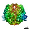 30237MC  7by0C M: map data used to model this data C: citing same article ( |
|---|---|
| Similar structure data | |
| EM raw data | EMPIAR-10571 (Title: The cryo-EM structure of the CENP-A nucleosome in complex with the phosphorylated CENP-C:: CENP-A nucleosome in complex with CENP-C motif (655-675) and CENP-N N-terminal domain (1-211) Data size: 1.3 TB Data #1: CENP-A nucleosome in complex with CENP-C motif (655-675) and CENP-N N-terminal domain (1-211) [micrographs - multiframe]) |


-Links
 PDBj
PDBj


- Assembly
| Deposited unit | 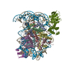
|
|---|---|
| 1 |
|
-Components
-Protein , 5 types, 10 molecules AEBFCGDHMN
| #1: Protein | Mass: 16372.217 Da / Num. of mol.: 2 Source method: isolated from a genetically manipulated source Details: chimeric protein, chicken histone H3 (aa 1-64)-chicken CENP-A (aa 52-131) Source: (gene. exp.)  #2: Protein | Mass: 11676.703 Da / Num. of mol.: 2 Source method: isolated from a genetically manipulated source Source: (gene. exp.) Homo sapiens (human) / Plasmid: pET15b / Production host: #3: Protein | Mass: 14447.825 Da / Num. of mol.: 2 Source method: isolated from a genetically manipulated source Source: (gene. exp.) Homo sapiens (human) / Gene: H2AC4, H2AFM, HIST1H2AB, H2AC8, H2AFA, HIST1H2AE / Plasmid: pET15b / Production host: #4: Protein | Mass: 14233.518 Da / Num. of mol.: 2 Source method: isolated from a genetically manipulated source Source: (gene. exp.) Homo sapiens (human) / Gene: H2BC21, H2BFQ, HIST2H2BE / Production host: #8: Protein | Mass: 71519.625 Da / Num. of mol.: 2 Source method: isolated from a genetically manipulated source Details: The fusion protein of N-terminal MBP, N-terminal domain of chicken CENP-N and C-terminal histidine tags Source: (gene. exp.) unidentified cloning vector (others), (gene. exp.) Gene: CENPN / Details (production host): pMAL_c2X / Production host: |
|---|
-DNA chain , 2 types, 2 molecules IJ
| #5: DNA chain | Mass: 44529.398 Da / Num. of mol.: 1 / Source method: obtained synthetically / Source: (synth.) synthetic construct (others) |
|---|---|
| #6: DNA chain | Mass: 44982.648 Da / Num. of mol.: 1 / Source method: obtained synthetically / Source: (synth.) synthetic construct (others) |
-Protein/peptide , 1 types, 2 molecules KL
| #7: Protein/peptide | Mass: 4932.840 Da / Num. of mol.: 2 / Source method: obtained synthetically / Source: (synth.) |
|---|
-Experimental details
-Experiment
| Experiment | Method: ELECTRON MICROSCOPY |
|---|---|
| EM experiment | Aggregation state: PARTICLE / 3D reconstruction method: single particle reconstruction |
- Sample preparation
| Component |
| ||||||||||||||||||||||||||||||||||||||||||||||||||||||
|---|---|---|---|---|---|---|---|---|---|---|---|---|---|---|---|---|---|---|---|---|---|---|---|---|---|---|---|---|---|---|---|---|---|---|---|---|---|---|---|---|---|---|---|---|---|---|---|---|---|---|---|---|---|---|---|
| Molecular weight | Value: 0.33 MDa / Experimental value: NO | ||||||||||||||||||||||||||||||||||||||||||||||||||||||
| Source (natural) |
| ||||||||||||||||||||||||||||||||||||||||||||||||||||||
| Buffer solution | pH: 7.5 | ||||||||||||||||||||||||||||||||||||||||||||||||||||||
| Buffer component |
| ||||||||||||||||||||||||||||||||||||||||||||||||||||||
| Specimen | Conc.: 1.3 mg/ml / Embedding applied: NO / Shadowing applied: NO / Staining applied: NO / Vitrification applied: YES | ||||||||||||||||||||||||||||||||||||||||||||||||||||||
| Specimen support | Details: Both side / Grid material: MOLYBDENUM / Grid mesh size: 200 divisions/in. / Grid type: Quantifoil R0.6/1 | ||||||||||||||||||||||||||||||||||||||||||||||||||||||
| Vitrification | Instrument: FEI VITROBOT MARK IV / Cryogen name: ETHANE / Humidity: 100 % / Chamber temperature: 277 K |
 Ectocarpus siliculosus (eukaryote)
- Electron microscopy imaging
| Microscopy | Model: JEOL CRYO ARM 200 |
|---|---|
| Electron gun | Electron source:  FIELD EMISSION GUN / Accelerating voltage: 200 kV / Illumination mode: FLOOD BEAM FIELD EMISSION GUN / Accelerating voltage: 200 kV / Illumination mode: FLOOD BEAM |
| Electron lens | Mode: BRIGHT FIELD / Nominal magnification: 50000 X / Calibrated magnification: 45454 X / Nominal defocus max: -3500 nm / Nominal defocus min: -1000 nm / Calibrated defocus min: -500 nm / Calibrated defocus max: -7000 nm / Cs: 1.4 mm / C2 aperture diameter: 150 µm / Alignment procedure: BASIC |
| Specimen holder | Cryogen: NITROGEN / Specimen holder model: JEOL CRYOSPECPORTER / Temperature (max): 80 K / Temperature (min): 80 K |
| Image recording | Average exposure time: 10 sec. / Electron dose: 1.2 e/Å2 / Detector mode: COUNTING / Film or detector model: GATAN K2 SUMMIT (4k x 4k) / Num. of grids imaged: 5 / Num. of real images: 5346 |
| Image scans | Sampling size: 5 µm / Width: 3838 / Height: 3710 / Movie frames/image: 50 / Used frames/image: 1-50 |
- Processing
| Software |
| ||||||||||||||||||||||||||||
|---|---|---|---|---|---|---|---|---|---|---|---|---|---|---|---|---|---|---|---|---|---|---|---|---|---|---|---|---|---|
| EM software |
| ||||||||||||||||||||||||||||
| CTF correction | Type: PHASE FLIPPING AND AMPLITUDE CORRECTION | ||||||||||||||||||||||||||||
| Particle selection | Num. of particles selected: 421635 | ||||||||||||||||||||||||||||
| Symmetry | Point symmetry: C2 (2 fold cyclic) | ||||||||||||||||||||||||||||
| 3D reconstruction | Resolution: 4.2 Å / Resolution method: FSC 0.143 CUT-OFF / Num. of particles: 118294 / Algorithm: FOURIER SPACE / Num. of class averages: 1 / Symmetry type: POINT | ||||||||||||||||||||||||||||
| Refinement | Cross valid method: NONE |
