 Movie
Movie Controller

[English] 日本語
 Yorodumi
- PDB-4jjn: Crystal structure of heterochromatin protein Sir3 in complex with... -
+ Open data

- Basic information
| Entry | Database: PDB / ID: 4jjn | ||||||
|---|---|---|---|---|---|---|---|
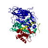
| Title | Crystal structure of heterochromatin protein Sir3 in complex with a silenced yeast nucleosome | ||||||
 Components Components |
| ||||||
 Keywords Keywords | DNA BINDING PROTEIN/DNA / BAH domain / silencing / DNA BINDING PROTEIN-DNA complex | ||||||
| Function / homology |  Function and homology information Function and homology informationsexual sporulation resulting in formation of a cellular spore / establishment of protein-containing complex localization to telomere / cupric reductase (NADH) activity / HATs acetylate histones / global genome nucleotide-excision repair / RNA polymerase I upstream activating factor complex / nuclear-transcribed mRNA catabolic process, non-stop decay / Condensation of Prophase Chromosomes / : / : ...sexual sporulation resulting in formation of a cellular spore / establishment of protein-containing complex localization to telomere / cupric reductase (NADH) activity / HATs acetylate histones / global genome nucleotide-excision repair / RNA polymerase I upstream activating factor complex / nuclear-transcribed mRNA catabolic process, non-stop decay / Condensation of Prophase Chromosomes / : / : / telomere tethering at nuclear periphery / : / Assembly of the ORC complex at the origin of replication / HDACs deacetylate histones / mitotic DNA replication checkpoint signaling / Oxidative Stress Induced Senescence / Recruitment and ATM-mediated phosphorylation of repair and signaling proteins at DNA double strand breaks / RMTs methylate histone arginines / chromatin silencing complex / silent mating-type cassette heterochromatin formation / SUMOylation of chromatin organization proteins / RNA Polymerase I Promoter Escape / negative regulation of DNA replication / positive regulation of transcription by RNA polymerase I / nucleolar large rRNA transcription by RNA polymerase I / Estrogen-dependent gene expression / rRNA transcription / DNA replication origin binding / intracellular copper ion homeostasis / DNA replication initiation / Ub-specific processing proteases / subtelomeric heterochromatin formation / nucleosome binding / heterochromatin / CENP-A containing nucleosome / aerobic respiration / double-strand break repair via nonhomologous end joining / structural constituent of chromatin / nucleosome / heterochromatin formation / nucleosome assembly / single-stranded DNA binding / chromatin organization / double-stranded DNA binding / nucleic acid binding / chromosome, telomeric region / protein heterodimerization activity / DNA repair / chromatin binding / regulation of DNA-templated transcription / nucleolus / mitochondrion / DNA binding / identical protein binding / nucleus Similarity search - Function | ||||||
| Biological species |  | ||||||
| Method |  X-RAY DIFFRACTION / SYNCHROTRON / MOLECULAR REPLACEMENT / Resolution: 3.09 Å X-RAY DIFFRACTION / SYNCHROTRON / MOLECULAR REPLACEMENT / Resolution: 3.09 Å | ||||||
 Authors Authors | Wang, F. / Li, G. / Mohammed, A. / Lu, C. / Currie, M. / Johnson, A. / Moazed, D. | ||||||
 Citation Citation | Journal: Proc.Natl.Acad.Sci.USA / Year: 2013 Title: Heterochromatin protein Sir3 induces contacts between the amino terminus of histone H4 and nucleosomal DNA. Authors: Wang, F. / Li, G. / Altaf, M. / Lu, C. / Currie, M.A. / Johnson, A. / Moazed, D. | ||||||
| History |
|
- Structure visualization
| Structure viewer | Molecule: MolmilJmol/JSmol |
|---|
- Downloads & links
-Download
| PDBx/mmCIF format | 4jjn.cif.gz | 776.1 KB | Display | PDBx/mmCIF format |
|---|---|---|---|---|
| PDB format | pdb4jjn.ent.gz | 628.8 KB | Display | PDB format |
| PDBx/mmJSON format | 4jjn.json.gz | Tree view | PDBx/mmJSON format | |
| Others |  Other downloads Other downloads |
-Validation report
| Arichive directory | https://data.pdbj.org/pub/pdb/validation_reports/jj/4jjnftp://data.pdbj.org/pub/pdb/validation_reports/jj/4jjn | HTTPS FTP |
|---|
-Related structure data
| Similar structure data |
|---|


-Links
 PDBj
PDBj


- Assembly
| Deposited unit | 
| ||||||||
|---|---|---|---|---|---|---|---|---|---|
| 1 |
| ||||||||
| Unit cell |
|
-Components
-Protein , 5 types, 10 molecules AEBFCGDHKL
| #1: Protein | Mass: 15259.811 Da / Num. of mol.: 2 Source method: isolated from a genetically manipulated source Source: (gene. exp.) Strain: ATCC 204508 / S288c Gene: HHT1, HHT2, Histone H3, N2749, SIN2, YBR010W, YBR0201, YNL031C Production host:  #2: Protein | Mass: 11264.194 Da / Num. of mol.: 2 Source method: isolated from a genetically manipulated source Source: (gene. exp.) Strain: ATCC 204508 / S288c Gene: HHF1, HHF2, Histone H4, N2752, YBR009C, YBR0122, YNL030W Production host: #3: Protein | Mass: 13881.980 Da / Num. of mol.: 2 Source method: isolated from a genetically manipulated source Source: (gene. exp.) Strain: ATCC 204508 / S288c / Gene: H2A2, Histone H2A.2, HTA2, YBL003C, YBL0103 / Production host: #4: Protein | Mass: 14133.145 Da / Num. of mol.: 2 Source method: isolated from a genetically manipulated source Source: (gene. exp.) Strain: ATCC 204508 / S288c / Gene: H2B2, Histone H2B.2, HTB2, YBL002W, YBL0104 / Production host: #5: Protein | Mass: 43655.582 Da / Num. of mol.: 2 / Mutation: D205N Source method: isolated from a genetically manipulated source Source: (gene. exp.) Strain: ATCC 204508 / S288c / Gene: CMT1, L9753.10, MAR2, SIR3, STE8, YLR442C / Plasmid: pET-Sumo / Production host: |
|---|
-DNA chain , 2 types, 2 molecules IJ
| #6: DNA chain | Mass: 45138.770 Da / Num. of mol.: 1 / Source method: obtained synthetically |
|---|---|
| #7: DNA chain | Mass: 45610.043 Da / Num. of mol.: 1 / Source method: obtained synthetically |
-Experimental details
-Experiment
| Experiment | Method: X-RAY DIFFRACTION / Number of used crystals: 1 |
|---|
- Sample preparation
| Crystal | Density Matthews: 3.01 Å3/Da / Density % sol: 59.09 % |
|---|---|
| Crystal grow | Temperature: 277 K / Method: vapor diffusion, sitting drop / pH: 7.5 Details: 0.05 M sodium cacodylate, 32% 2-methyl-2,4-pentanediol (MPD), pH 7.5, VAPOR DIFFUSION, SITTING DROP, temperature 277K |
-Data collection
| Diffraction | Mean temperature: 100 K | |||||||||||||||||||||||||||||||||||||||||||||||||||||||||||||||||||||||||||||
|---|---|---|---|---|---|---|---|---|---|---|---|---|---|---|---|---|---|---|---|---|---|---|---|---|---|---|---|---|---|---|---|---|---|---|---|---|---|---|---|---|---|---|---|---|---|---|---|---|---|---|---|---|---|---|---|---|---|---|---|---|---|---|---|---|---|---|---|---|---|---|---|---|---|---|---|---|---|---|
| Diffraction source | Source: SYNCHROTRON / Site: APS  / Beamline: 24-ID-C / Wavelength: 0.9792 Å / Beamline: 24-ID-C / Wavelength: 0.9792 Å | |||||||||||||||||||||||||||||||||||||||||||||||||||||||||||||||||||||||||||||
| Detector | Type: PSI PILATUS 6M / Detector: PIXEL / Date: Aug 15, 2012 | |||||||||||||||||||||||||||||||||||||||||||||||||||||||||||||||||||||||||||||
| Radiation | Monochromator: SAGITALLY FOCUSED Si(111) / Protocol: SINGLE WAVELENGTH / Monochromatic (M) / Laue (L): M / Scattering type: x-ray | |||||||||||||||||||||||||||||||||||||||||||||||||||||||||||||||||||||||||||||
| Radiation wavelength | Wavelength: 0.9792 Å / Relative weight: 1 | |||||||||||||||||||||||||||||||||||||||||||||||||||||||||||||||||||||||||||||
| Reflection | Resolution: 3.09→139.09 Å / Num. all: 61627 / Num. obs: 60025 / % possible obs: 97.4 % / Observed criterion σ(F): -3 / Observed criterion σ(I): -3 / Redundancy: 2.9 % / Rmerge(I) obs: 0.039 / Rsym value: 0.039 / Net I/σ(I): 14 | |||||||||||||||||||||||||||||||||||||||||||||||||||||||||||||||||||||||||||||
| Reflection shell |
|
- Processing
| Software |
| |||||||||||||||||||||||||||||||||||||||||||||||||||||||||||||||||||||||||||||||||||||||||||||||||||||||||||||||||||||||||||||||||||||||||||||||||||||||||||||||||
|---|---|---|---|---|---|---|---|---|---|---|---|---|---|---|---|---|---|---|---|---|---|---|---|---|---|---|---|---|---|---|---|---|---|---|---|---|---|---|---|---|---|---|---|---|---|---|---|---|---|---|---|---|---|---|---|---|---|---|---|---|---|---|---|---|---|---|---|---|---|---|---|---|---|---|---|---|---|---|---|---|---|---|---|---|---|---|---|---|---|---|---|---|---|---|---|---|---|---|---|---|---|---|---|---|---|---|---|---|---|---|---|---|---|---|---|---|---|---|---|---|---|---|---|---|---|---|---|---|---|---|---|---|---|---|---|---|---|---|---|---|---|---|---|---|---|---|---|---|---|---|---|---|---|---|---|---|---|---|---|---|---|---|
| Refinement | Method to determine structure: MOLECULAR REPLACEMENT / Resolution: 3.09→85.45 Å / SU ML: 0.43 / σ(F): 1.35 / Phase error: 30.27 / Stereochemistry target values: ML
| |||||||||||||||||||||||||||||||||||||||||||||||||||||||||||||||||||||||||||||||||||||||||||||||||||||||||||||||||||||||||||||||||||||||||||||||||||||||||||||||||
| Solvent computation | Shrinkage radii: 0.9 Å / VDW probe radii: 1.11 Å / Solvent model: FLAT BULK SOLVENT MODEL | |||||||||||||||||||||||||||||||||||||||||||||||||||||||||||||||||||||||||||||||||||||||||||||||||||||||||||||||||||||||||||||||||||||||||||||||||||||||||||||||||
| Displacement parameters | Biso mean: 78.24 Å2 | |||||||||||||||||||||||||||||||||||||||||||||||||||||||||||||||||||||||||||||||||||||||||||||||||||||||||||||||||||||||||||||||||||||||||||||||||||||||||||||||||
| Refinement step | Cycle: LAST / Resolution: 3.09→85.45 Å
| |||||||||||||||||||||||||||||||||||||||||||||||||||||||||||||||||||||||||||||||||||||||||||||||||||||||||||||||||||||||||||||||||||||||||||||||||||||||||||||||||
| Refine LS restraints |
| |||||||||||||||||||||||||||||||||||||||||||||||||||||||||||||||||||||||||||||||||||||||||||||||||||||||||||||||||||||||||||||||||||||||||||||||||||||||||||||||||
| LS refinement shell |
| |||||||||||||||||||||||||||||||||||||||||||||||||||||||||||||||||||||||||||||||||||||||||||||||||||||||||||||||||||||||||||||||||||||||||||||||||||||||||||||||||
| Refinement TLS params. | Method: refined / Origin x: 11.8658 Å / Origin y: -20.1545 Å / Origin z: 24.5817 Å
| |||||||||||||||||||||||||||||||||||||||||||||||||||||||||||||||||||||||||||||||||||||||||||||||||||||||||||||||||||||||||||||||||||||||||||||||||||||||||||||||||
| Refinement TLS group | Selection details: ALL |
