: / meiotic recombination checkpoint signaling / [histone H3]-lysine79 N-trimethyltransferase / histone H3K79 trimethyltransferase activity / histone H3K79 methyltransferase activity / global genome nucleotide-excision repair / negative regulation of heterochromatin formation / DNA damage tolerance / mitotic intra-S DNA damage checkpoint signaling / recombinational repair ...: / meiotic recombination checkpoint signaling / [histone H3]-lysine79 N-trimethyltransferase / histone H3K79 trimethyltransferase activity / histone H3K79 methyltransferase activity / global genome nucleotide-excision repair / negative regulation of heterochromatin formation / DNA damage tolerance / mitotic intra-S DNA damage checkpoint signaling / recombinational repair / subtelomeric heterochromatin formation / mitotic G1 DNA damage checkpoint signaling / DNA damage checkpoint signaling / Maturation of protein E / Maturation of protein E / ER Quality Control Compartment (ERQC) / Myoclonic epilepsy of Lafora / FLT3 signaling by CBL mutants / IRAK2 mediated activation of TAK1 complex / Alpha-protein kinase 1 signaling pathway / Glycogen synthesis / IRAK1 recruits IKK complex / IRAK1 recruits IKK complex upon TLR7/8 or 9 stimulation / Prevention of phagosomal-lysosomal fusion / Endosomal Sorting Complex Required For Transport (ESCRT) / Membrane binding and targetting of GAG proteins / Regulation of TBK1, IKKε (IKBKE)-mediated activation of IRF3, IRF7 / Negative regulation of FLT3 / PTK6 Regulates RTKs and Their Effectors AKT1 and DOK1 / Regulation of TBK1, IKKε-mediated activation of IRF3, IRF7 upon TLR3 ligation / IRAK2 mediated activation of TAK1 complex upon TLR7/8 or 9 stimulation / Constitutive Signaling by NOTCH1 HD Domain Mutants / NOTCH2 Activation and Transmission of Signal to the Nucleus / TICAM1,TRAF6-dependent induction of TAK1 complex / TICAM1-dependent activation of IRF3/IRF7 / APC/C:Cdc20 mediated degradation of Cyclin B / Downregulation of ERBB4 signaling / APC-Cdc20 mediated degradation of Nek2A / Regulation of FZD by ubiquitination / p75NTR recruits signalling complexes / InlA-mediated entry of Listeria monocytogenes into host cells / TRAF6 mediated IRF7 activation in TLR7/8 or 9 signaling / NF-kB is activated and signals survival / TRAF6-mediated induction of TAK1 complex within TLR4 complex / Regulation of pyruvate metabolism / Pexophagy / Downregulation of ERBB2:ERBB3 signaling / Regulation of innate immune responses to cytosolic DNA / NRIF signals cell death from the nucleus / Regulation of PTEN localization / VLDLR internalisation and degradation / Activated NOTCH1 Transmits Signal to the Nucleus / Synthesis of active ubiquitin: roles of E1 and E2 enzymes / Translesion synthesis by REV1 / TICAM1, RIP1-mediated IKK complex recruitment / Regulation of BACH1 activity / Translesion synthesis by POLK / JNK (c-Jun kinases) phosphorylation and activation mediated by activated human TAK1 / InlB-mediated entry of Listeria monocytogenes into host cell / MAP3K8 (TPL2)-dependent MAPK1/3 activation / Activation of IRF3, IRF7 mediated by TBK1, IKKε (IKBKE) / Downregulation of TGF-beta receptor signaling / Translesion synthesis by POLI / Josephin domain DUBs / Gap-filling DNA repair synthesis and ligation in GG-NER / IKK complex recruitment mediated by RIP1 / PINK1-PRKN Mediated Mitophagy / TGF-beta receptor signaling in EMT (epithelial to mesenchymal transition) / TNFR1-induced NF-kappa-B signaling pathway / Regulation of activated PAK-2p34 by proteasome mediated degradation / TCF dependent signaling in response to WNT / Regulation of NF-kappa B signaling / activated TAK1 mediates p38 MAPK activation / nucleotide-excision repair / Autodegradation of Cdh1 by Cdh1:APC/C / APC/C:Cdc20 mediated degradation of Securin / NOTCH3 Activation and Transmission of Signal to the Nucleus / Regulation of signaling by CBL / Negative regulators of DDX58/IFIH1 signaling / N-glycan trimming in the ER and Calnexin/Calreticulin cycle / Asymmetric localization of PCP proteins / Fanconi Anemia Pathway / Negative regulation of FGFR3 signaling / Ubiquitin-dependent degradation of Cyclin D / Peroxisomal protein import / Deactivation of the beta-catenin transactivating complex / SCF-beta-TrCP mediated degradation of Emi1 / NIK-->noncanonical NF-kB signaling / Stabilization of p53 / AUF1 (hnRNP D0) binds and destabilizes mRNA / TNFR2 non-canonical NF-kB pathway / Negative regulation of FGFR2 signaling / Negative regulation of FGFR4 signaling / Downregulation of SMAD2/3:SMAD4 transcriptional activity / Negative regulation of FGFR1 signaling / Termination of translesion DNA synthesis / Assembly of the pre-replicative complex / Vpu mediated degradation of CD4 / EGFR downregulation / Regulation of TNFR1 signaling Similarity search - Function
National Institutes of Health/National Institute of General Medical Sciences (NIH/NIGMS)
5R01GM115882-03
United States
Citation
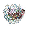
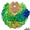
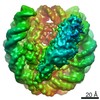
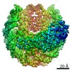
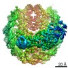
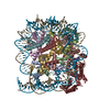
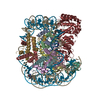
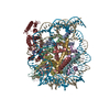
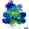
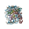
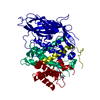
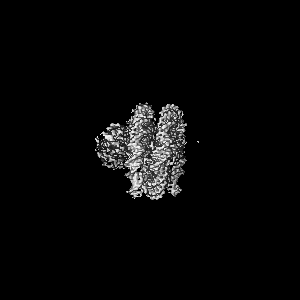
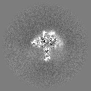
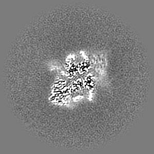
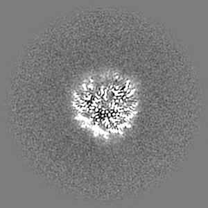
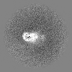
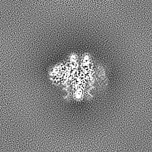
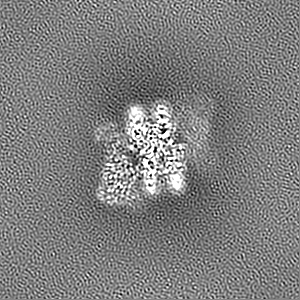
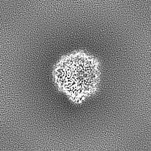
Journal: Science / Year: 2021 Title: Regulation of the Dot1 histone H3K79 methyltransferase by histone H4K16 acetylation. Authors: Marco Igor Valencia-Sánchez / Pablo De Ioannes / Miao Wang / David M Truong / Rachel Lee / Jean-Paul Armache / Jef D Boeke / Karim-Jean Armache / Abstract: Dot1 (disruptor of telomeric silencing-1), the histone H3 lysine 79 (H3K79) methyltransferase, is conserved throughout evolution, and its deregulation is found in human leukemias. Here, we provide ...Dot1 (disruptor of telomeric silencing-1), the histone H3 lysine 79 (H3K79) methyltransferase, is conserved throughout evolution, and its deregulation is found in human leukemias. Here, we provide evidence that acetylation of histone H4 allosterically stimulates yeast Dot1 in a manner distinct from but coordinating with histone H2B ubiquitination (H2BUb). We further demonstrate that this stimulatory effect is specific to acetylation of lysine 16 (H4K16ac), a modification central to chromatin structure. We provide a mechanism of this histone cross-talk and show that H4K16ac and H2BUb play crucial roles in H3K79 di- and trimethylation in vitro and in vivo. These data reveal mechanisms that control H3K79 methylation and demonstrate how H4K16ac, H3K79me, and H2BUb function together to regulate gene transcription and gene silencing to ensure optimal maintenance and propagation of an epigenetic state.
History
Deposition
Sep 21, 2020
-
Header (metadata) release
Feb 10, 2021
-
Map release
Feb 10, 2021
-
Update
Oct 23, 2024
-
Current status
Oct 23, 2024
Processing site: RCSB / Status: Released
-
Structure visualization
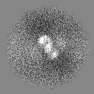
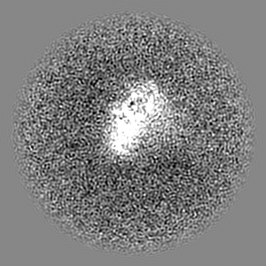
Movie
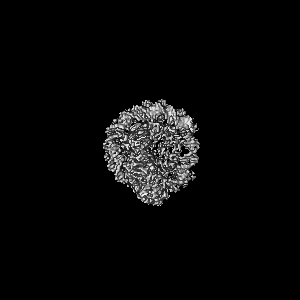
Surface view with section colored by density value
Model: Quantifoil R1.2/1.3 / Material: GOLD / Mesh: 400 / Support film - Material: CARBON / Support film - topology: HOLEY / Pretreatment - Type: GLOW DISCHARGE / Pretreatment - Time: 30 sec.
Vitrification
Cryogen name: ETHANE / Chamber humidity: 100 % / Chamber temperature: 297 K / Instrument: FEI VITROBOT MARK IV
-
Electron microscopy
Microscope
FEI TITAN KRIOS
Specialist optics
Energy filter - Slit width: 20 eV
Image recording
Film or detector model: GATAN K2 SUMMIT (4k x 4k) / Detector mode: COUNTING / Number grids imaged: 1 / Number real images: 1809 / Average exposure time: 10.0 sec. / Average electron dose: 74.5 e/Å2
Electron beam
Acceleration voltage: 300 kV / Electron source: FIELD EMISSION GUN
Electron optics
Illumination mode: OTHER / Imaging mode: BRIGHT FIELD / Cs: 2.7 mm / Nominal defocus max: 2.4 µm / Nominal defocus min: 1.2 µm / Nominal magnification: 130000
In the structure databanks used in Yorodumi, some data are registered as the other names, "COVID-19 virus" and "2019-nCoV". Here are the details of the virus and the list of structure data.
Jan 31, 2019. EMDB accession codes are about to change! (news from PDBe EMDB page)
EMDB accession codes are about to change! (news from PDBe EMDB page)
The allocation of 4 digits for EMDB accession codes will soon come to an end. Whilst these codes will remain in use, new EMDB accession codes will include an additional digit and will expand incrementally as the available range of codes is exhausted. The current 4-digit format prefixed with “EMD-” (i.e. EMD-XXXX) will advance to a 5-digit format (i.e. EMD-XXXXX), and so on. It is currently estimated that the 4-digit codes will be depleted around Spring 2019, at which point the 5-digit format will come into force.
The EM Navigator/Yorodumi systems omit the EMD- prefix.
Related info.:Q: What is EMD? / ID/Accession-code notation in Yorodumi/EM Navigator
Yorodumi is a browser for structure data from EMDB, PDB, SASBDB, etc.
This page is also the successor to EM Navigator detail page, and also detail information page/front-end page for Omokage search.
The word "yorodu" (or yorozu) is an old Japanese word meaning "ten thousand". "mi" (miru) is to see.
Related info.:EMDB / PDB / SASBDB / Comparison of 3 databanks / Yorodumi Search / Aug 31, 2016. New EM Navigator & Yorodumi / Yorodumi Papers / Jmol/JSmol / Function and homology information / Changes in new EM Navigator and Yorodumi
 Movie
Movie Controller


 Open data
 Basic information
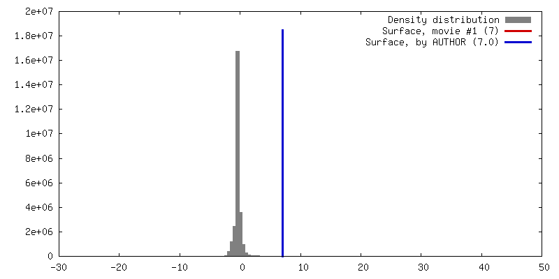
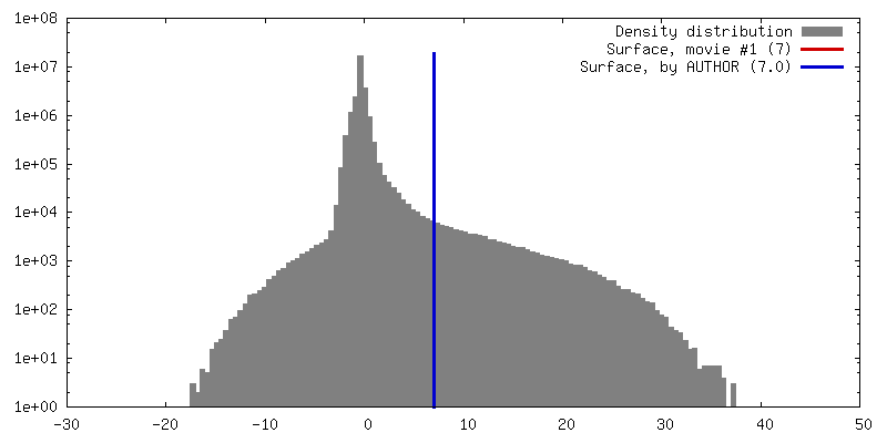
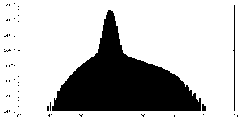
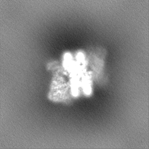
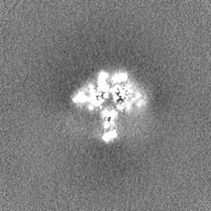
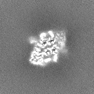
 Map data
 Sample
 Keywords
 Function and homology information
 Homo sapiens (human) / Synthetic construct (others) / synthetic construct (others)
 Authors
 United States, 2 items
  Citation
 Structure visualization
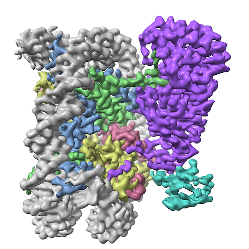
 Downloads & links
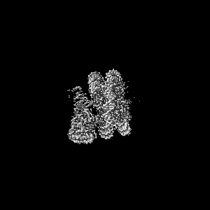
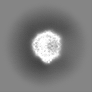
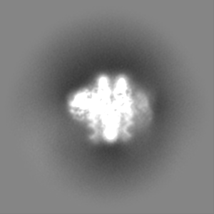
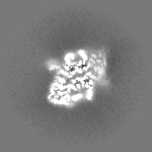
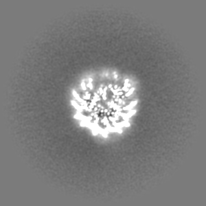
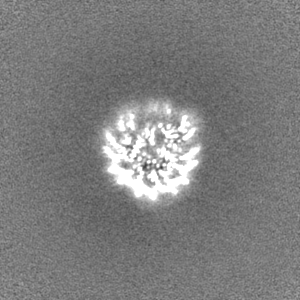
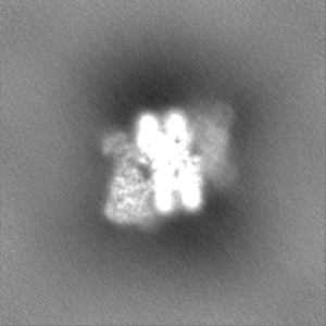
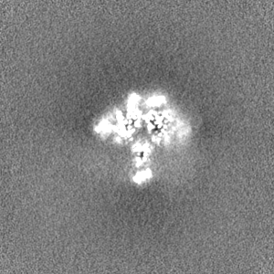
 emd_22692.png
 http://ftp.pdbj.org/pub/emdb/structures/EMD-22692


 Z (Sec.)
 Y (Row.)
 X (Col.)


 Sample components

 Processing
 Electron microscopy
 FIELD EMISSION GUN