 Movie
Movie Controller


+ Open data

- Basic information
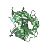
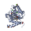
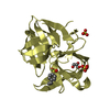
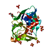
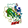
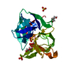
| Entry | Database: PDB / ID: 7bf0 | ||||||
|---|---|---|---|---|---|---|---|
| Title | Zn-bound domain 3 of CDCA1 in complex with carbon dioxide | ||||||
 Components Components | Cadmium-specific carbonic anhydrase | ||||||
 Keywords Keywords | LYASE / CARBON DIOXIDE / CARBONIC ANHYDRASE / ZINC-BOUND / MARINE DIATOM | ||||||
| Function / homology | Cadmium carbonic anhydrase repeat / Cadmium carbonic anhydrase repeat / metal ion binding / CARBON DIOXIDE / Cadmium-specific carbonic anhydrase Function and homology information Function and homology information | ||||||
| Biological species |  Thalassiosira weissflogii (Diatom) Thalassiosira weissflogii (Diatom) | ||||||
| Method |  X-RAY DIFFRACTION / SYNCHROTRON / MOLECULAR REPLACEMENT / Resolution: 1.6 Å X-RAY DIFFRACTION / SYNCHROTRON / MOLECULAR REPLACEMENT / Resolution: 1.6 Å | ||||||
 Authors Authors | Alterio, V. / De Simone, G. | ||||||
 Citation Citation | Journal: Comput Struct Biotechnol J / Year: 2021 Title: Zeta-carbonic anhydrases show CS 2 hydrolase activity: A new metabolic carbon acquisition pathway in diatoms? Authors: Alterio, V. / Langella, E. / Buonanno, M. / Esposito, D. / Nocentini, A. / Berrino, E. / Bua, S. / Polentarutti, M. / Supuran, C.T. / Monti, S.M. / De Simone, G. | ||||||
| History |
|
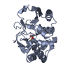
- Structure visualization
| Structure viewer | Molecule: MolmilJmol/JSmol |
|---|
- Downloads & links
-Download
| PDBx/mmCIF format | 7bf0.cif.gz | 98 KB | Display | PDBx/mmCIF format |
|---|---|---|---|---|
| PDB format | pdb7bf0.ent.gz | 73.5 KB | Display | PDB format |
| PDBx/mmJSON format | 7bf0.json.gz | Tree view | PDBx/mmJSON format | |
| Others |  Other downloads Other downloads |
-Validation report
| Summary document | 7bf0_validation.pdf.gz | 327.2 KB | Display | wwPDB validaton report |
|---|---|---|---|---|
| Full document | 7bf0_full_validation.pdf.gz | 328.7 KB | Display | |
| Data in XML | 7bf0_validation.xml.gz | 18.2 KB | Display | |
| Data in CIF | 7bf0_validation.cif.gz | 26 KB | Display | |
| Arichive directory | https://data.pdbj.org/pub/pdb/validation_reports/bf/7bf0ftp://data.pdbj.org/pub/pdb/validation_reports/bf/7bf0 | HTTPS FTP |
-Related structure data
| Related structure data | 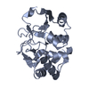 7bezC 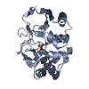 3uk8S S: Starting model for refinement C: citing same article ( |
|---|---|
| Similar structure data |

-Links
 PDBj
PDBj

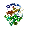
- Assembly
| Deposited unit | 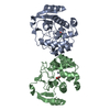
| ||||||||
|---|---|---|---|---|---|---|---|---|---|
| 1 | 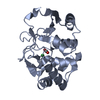
| ||||||||
| 2 | 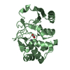
| ||||||||
| Unit cell |
|
-Components
| #1: Protein | Mass: 24826.391 Da / Num. of mol.: 2 Source method: isolated from a genetically manipulated source Details: MARINE DIATOM / Source: (gene. exp.) Thalassiosira weissflogii (Diatom) / Gene: cdca1 / Plasmid: PET15B / Production host:  #2: Chemical |   Mass: 65.409 Da / Num. of mol.: 2 / Source method: obtained synthetically / Formula: Zn Mass: 65.409 Da / Num. of mol.: 2 / Source method: obtained synthetically / Formula: Zn#3: Chemical | ChemComp-CL / |   Mass: 35.453 Da / Num. of mol.: 1 / Source method: obtained synthetically / Formula: Cl Mass: 35.453 Da / Num. of mol.: 1 / Source method: obtained synthetically / Formula: Cl#4: Chemical |   Mass: 44.010 Da / Num. of mol.: 2 / Source method: obtained synthetically / Formula: CO2 / Feature type: SUBJECT OF INVESTIGATION Mass: 44.010 Da / Num. of mol.: 2 / Source method: obtained synthetically / Formula: CO2 / Feature type: SUBJECT OF INVESTIGATION#5: Water | ChemComp-HOH / |  Mass: 18.015 Da / Num. of mol.: 171 / Source method: isolated from a natural source / Formula: H2O Mass: 18.015 Da / Num. of mol.: 171 / Source method: isolated from a natural source / Formula: H2OHas ligand of interest | Y | |
|---|
-Experimental details
-Experiment
| Experiment | Method: X-RAY DIFFRACTION / Number of used crystals: 1 |
|---|
- Sample preparation
| Crystal | Density Matthews: 2.62 Å3/Da / Density % sol: 53 % |
|---|---|
| Crystal grow | Temperature: 293 K / Method: vapor diffusion, hanging drop / pH: 5.9 Details: 28% (w/v) PEG2000, 20 mM lithium sulfate, 100 mM sodium citrate |
-Data collection
| Diffraction | Mean temperature: 100 K / Serial crystal experiment: N | ||||||||||||||||||||||||||||||||||||||||||||||||||||||||||||||||||||||||||||||||||||||||||||||||||||||||||||||||||||||||||||||||||||||||||||||||||||||||||||||||||||||||||||||||||||||||||||||||||||||||||||||||||
|---|---|---|---|---|---|---|---|---|---|---|---|---|---|---|---|---|---|---|---|---|---|---|---|---|---|---|---|---|---|---|---|---|---|---|---|---|---|---|---|---|---|---|---|---|---|---|---|---|---|---|---|---|---|---|---|---|---|---|---|---|---|---|---|---|---|---|---|---|---|---|---|---|---|---|---|---|---|---|---|---|---|---|---|---|---|---|---|---|---|---|---|---|---|---|---|---|---|---|---|---|---|---|---|---|---|---|---|---|---|---|---|---|---|---|---|---|---|---|---|---|---|---|---|---|---|---|---|---|---|---|---|---|---|---|---|---|---|---|---|---|---|---|---|---|---|---|---|---|---|---|---|---|---|---|---|---|---|---|---|---|---|---|---|---|---|---|---|---|---|---|---|---|---|---|---|---|---|---|---|---|---|---|---|---|---|---|---|---|---|---|---|---|---|---|---|---|---|---|---|---|---|---|---|---|---|---|---|---|---|---|---|
| Diffraction source | Source: SYNCHROTRON / Site: ELETTRA  / Beamline: 5.2R / Wavelength: 0.999995 Å / Beamline: 5.2R / Wavelength: 0.999995 Å | ||||||||||||||||||||||||||||||||||||||||||||||||||||||||||||||||||||||||||||||||||||||||||||||||||||||||||||||||||||||||||||||||||||||||||||||||||||||||||||||||||||||||||||||||||||||||||||||||||||||||||||||||||
| Detector | Type: DECTRIS PILATUS 2M / Detector: PIXEL / Date: May 10, 2012 | ||||||||||||||||||||||||||||||||||||||||||||||||||||||||||||||||||||||||||||||||||||||||||||||||||||||||||||||||||||||||||||||||||||||||||||||||||||||||||||||||||||||||||||||||||||||||||||||||||||||||||||||||||
| Radiation | Protocol: SINGLE WAVELENGTH / Monochromatic (M) / Laue (L): M / Scattering type: x-ray | ||||||||||||||||||||||||||||||||||||||||||||||||||||||||||||||||||||||||||||||||||||||||||||||||||||||||||||||||||||||||||||||||||||||||||||||||||||||||||||||||||||||||||||||||||||||||||||||||||||||||||||||||||
| Radiation wavelength | Wavelength: 0.999995 Å / Relative weight: 1 | ||||||||||||||||||||||||||||||||||||||||||||||||||||||||||||||||||||||||||||||||||||||||||||||||||||||||||||||||||||||||||||||||||||||||||||||||||||||||||||||||||||||||||||||||||||||||||||||||||||||||||||||||||
| Reflection | Resolution: 1.6→50 Å / Num. obs: 61450 / % possible obs: 97 % / Redundancy: 11.6 % / CC1/2: 0.999 / Rmerge(I) obs: 0.066 / Rpim(I) all: 0.019 / Rrim(I) all: 0.068 / Χ2: 1.039 / Net I/av σ(I): 35.8 / Net I/σ(I): 14.6 | ||||||||||||||||||||||||||||||||||||||||||||||||||||||||||||||||||||||||||||||||||||||||||||||||||||||||||||||||||||||||||||||||||||||||||||||||||||||||||||||||||||||||||||||||||||||||||||||||||||||||||||||||||
| Reflection shell | Diffraction-ID: 1
|
- Processing
| Software |
| ||||||||||||||||||||||||||||
|---|---|---|---|---|---|---|---|---|---|---|---|---|---|---|---|---|---|---|---|---|---|---|---|---|---|---|---|---|---|
| Refinement | Method to determine structure: MOLECULAR REPLACEMENT Starting model: 3UK8 Resolution: 1.6→50 Å / Cross valid method: FREE R-VALUE / σ(F): 0
| ||||||||||||||||||||||||||||
| Displacement parameters | Biso max: 60.81 Å2 / Biso mean: 26.3624 Å2 / Biso min: 12.08 Å2
| ||||||||||||||||||||||||||||
| Refinement step | Cycle: final / Resolution: 1.6→50 Å
| ||||||||||||||||||||||||||||
| Refine LS restraints |
| ||||||||||||||||||||||||||||
| LS refinement shell |
|
