 Movie
Movie Controller

[English] 日本語
 Yorodumi
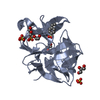
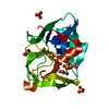
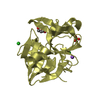
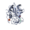
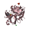
- PDB-6qoy: Crystal structure of L1 protease Lysobacter sp. XL1 in complex wi... -
+ Open data

- Basic information
| Entry | Database: PDB / ID: 6qoy | ||||||
|---|---|---|---|---|---|---|---|
| Title | Crystal structure of L1 protease Lysobacter sp. XL1 in complex with AEBSF | ||||||
 Components Components | Lytic endopeptidase preproenzyme | ||||||
 Keywords Keywords | HYDROLASE / Bacteriolytic protease L1 / Lysobacter sp. XL1 / Crystals / AEBSF | ||||||
| Function / homology |  Function and homology information Function and homology information | ||||||
| Biological species | Lysobacter sp. | ||||||
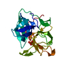
| Method |  X-RAY DIFFRACTION / SYNCHROTRON / MOLECULAR REPLACEMENT / Resolution: 1.9 Å X-RAY DIFFRACTION / SYNCHROTRON / MOLECULAR REPLACEMENT / Resolution: 1.9 Å | ||||||
 Authors Authors | Gabdulkhakov, A. / Tishchenko, S. / Kudryakova, I. / Afoshin, A. / Vasilyeva, N. | ||||||
 Citation Citation | Journal: Process Biochem / Year: 2019 Title: Serine bacteriolytic protease L1 of Lysobacter sp. XL1 complexed with protease inhibitor AEBSF: features of interaction Authors: Kudryakova, I. / Gabdulkhakov, A. / Tishchenko, S. / Afoshin, A. / Vasilyeva, N. | ||||||
| History |
|
- Structure visualization
| Structure viewer | Molecule: MolmilJmol/JSmol |
|---|
- Downloads & links
-Download
| PDBx/mmCIF format | 6qoy.cif.gz | 165.4 KB | Display | PDBx/mmCIF format |
|---|---|---|---|---|
| PDB format | pdb6qoy.ent.gz | 130.6 KB | Display | PDB format |
| PDBx/mmJSON format | 6qoy.json.gz | Tree view | PDBx/mmJSON format | |
| Others |  Other downloads Other downloads |
-Validation report
| Arichive directory | https://data.pdbj.org/pub/pdb/validation_reports/qo/6qoyftp://data.pdbj.org/pub/pdb/validation_reports/qo/6qoy | HTTPS FTP |
|---|
-Related structure data
| Related structure data |  5mrrS S: Starting model for refinement |
|---|---|
| Similar structure data |


-Links
 PDBj
PDBj- Assembly
| Deposited unit | 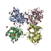
| ||||||||
|---|---|---|---|---|---|---|---|---|---|
| 1 | 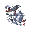
| ||||||||
| 2 | 
| ||||||||
| 3 | 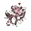
| ||||||||
| 4 | 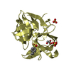
| ||||||||
| Unit cell |
|
-Components
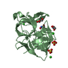
-Protein , 1 types, 4 molecules ABCD
| #1: Protein | Mass: 19848.051 Da / Num. of mol.: 4 Source method: isolated from a genetically manipulated source Source: (gene. exp.)  Lysobacter sp. (strain XL1) (bacteria) / Strain: XL1 / Gene: alpA / Plasmid: PAPROZ / Production host: Lysobacter sp. (strain XL1) (bacteria) / Strain: XL1 / Gene: alpA / Plasmid: PAPROZ / Production host: |
|---|
-Non-polymers , 8 types, 337 molecules 
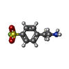


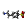


| #2: Chemical | ChemComp-SO4 /  Mass: 96.063 Da / Num. of mol.: 8 / Source method: obtained synthetically / Formula: SO4 Mass: 96.063 Da / Num. of mol.: 8 / Source method: obtained synthetically / Formula: SO4#3: Chemical | ChemComp-AES /  Mass: 203.234 Da / Num. of mol.: 4 / Source method: obtained synthetically / Formula: C8H10FNO2S / Comment: protease inhibitor*YM Mass: 203.234 Da / Num. of mol.: 4 / Source method: obtained synthetically / Formula: C8H10FNO2S / Comment: protease inhibitor*YM#4: Chemical | ChemComp-GOL /  Mass: 92.094 Da / Num. of mol.: 5 / Source method: obtained synthetically / Formula: C3H8O3 Mass: 92.094 Da / Num. of mol.: 5 / Source method: obtained synthetically / Formula: C3H8O3#5: Chemical | ChemComp-PGE / |  Mass: 150.173 Da / Num. of mol.: 1 / Source method: obtained synthetically / Formula: C6H14O4 Mass: 150.173 Da / Num. of mol.: 1 / Source method: obtained synthetically / Formula: C6H14O4#6: Chemical |  Mass: 201.243 Da / Num. of mol.: 3 / Source method: obtained synthetically / Formula: C8H11NO3S Mass: 201.243 Da / Num. of mol.: 3 / Source method: obtained synthetically / Formula: C8H11NO3S#7: Chemical |  Mass: 35.453 Da / Num. of mol.: 2 / Source method: obtained synthetically / Formula: Cl Mass: 35.453 Da / Num. of mol.: 2 / Source method: obtained synthetically / Formula: Cl#8: Chemical |  Mass: 106.120 Da / Num. of mol.: 3 / Source method: obtained synthetically / Formula: C4H10O3 Mass: 106.120 Da / Num. of mol.: 3 / Source method: obtained synthetically / Formula: C4H10O3#9: Water | ChemComp-HOH / | Mass: 18.015 Da / Num. of mol.: 311 / Source method: isolated from a natural source / Formula: H2O |
|---|
-Details
| Has protein modification | Y |
|---|
-Experimental details
-Experiment
| Experiment | Method: X-RAY DIFFRACTION / Number of used crystals: 1 |
|---|
- Sample preparation
| Crystal | Density Matthews: 2.52 Å3/Da / Density % sol: 51.24 % |
|---|---|
| Crystal grow | Temperature: 295 K / Method: vapor diffusion, hanging drop / pH: 6.5 / Details: 1,4M Lithium sulphate, 0,1M BisTris, |
-Data collection
| Diffraction | Mean temperature: 100 K / Serial crystal experiment: N |
|---|---|
| Diffraction source | Source: SYNCHROTRON / Site: BESSY  / Beamline: 14.1 / Wavelength: 0.96863 Å / Beamline: 14.1 / Wavelength: 0.96863 Å |
| Detector | Type: DECTRIS PILATUS3 S 6M / Detector: PIXEL / Date: Jul 10, 2015 |
| Radiation | Protocol: SINGLE WAVELENGTH / Monochromatic (M) / Laue (L): M / Scattering type: x-ray |
| Radiation wavelength | Wavelength: 0.96863 Å / Relative weight: 1 |
| Reflection | Resolution: 1.86→50 Å / Num. obs: 65564 / % possible obs: 99.4 % / Redundancy: 4.8 % / Rmerge(I) obs: 0.14 / Net I/σ(I): 8.13 |
| Reflection shell | Resolution: 1.86→1.97 Å / Rmerge(I) obs: 0.8 / Num. unique all: 10356 |
- Processing
| Software |
| |||||||||||||||||||||||||||||||||||||||||||||||||||||||||||||||||||||||||||||||||||||||||||||||||||||||||||||||||||||||||||||||||||||||||||||||||||||||||||||||||||||||||||||||||||||||||||||||||||||||||||||||||||||||||
|---|---|---|---|---|---|---|---|---|---|---|---|---|---|---|---|---|---|---|---|---|---|---|---|---|---|---|---|---|---|---|---|---|---|---|---|---|---|---|---|---|---|---|---|---|---|---|---|---|---|---|---|---|---|---|---|---|---|---|---|---|---|---|---|---|---|---|---|---|---|---|---|---|---|---|---|---|---|---|---|---|---|---|---|---|---|---|---|---|---|---|---|---|---|---|---|---|---|---|---|---|---|---|---|---|---|---|---|---|---|---|---|---|---|---|---|---|---|---|---|---|---|---|---|---|---|---|---|---|---|---|---|---|---|---|---|---|---|---|---|---|---|---|---|---|---|---|---|---|---|---|---|---|---|---|---|---|---|---|---|---|---|---|---|---|---|---|---|---|---|---|---|---|---|---|---|---|---|---|---|---|---|---|---|---|---|---|---|---|---|---|---|---|---|---|---|---|---|---|---|---|---|---|---|---|---|---|---|---|---|---|---|---|---|---|---|---|---|---|
| Refinement | Method to determine structure: MOLECULAR REPLACEMENT Starting model: 5MRR Resolution: 1.9→48.207 Å / SU ML: 0.27 / Cross valid method: FREE R-VALUE / σ(F): 1.29 / Phase error: 26.51
| |||||||||||||||||||||||||||||||||||||||||||||||||||||||||||||||||||||||||||||||||||||||||||||||||||||||||||||||||||||||||||||||||||||||||||||||||||||||||||||||||||||||||||||||||||||||||||||||||||||||||||||||||||||||||
| Solvent computation | Shrinkage radii: 0.9 Å / VDW probe radii: 1.11 Å | |||||||||||||||||||||||||||||||||||||||||||||||||||||||||||||||||||||||||||||||||||||||||||||||||||||||||||||||||||||||||||||||||||||||||||||||||||||||||||||||||||||||||||||||||||||||||||||||||||||||||||||||||||||||||
| Refinement step | Cycle: LAST / Resolution: 1.9→48.207 Å
| |||||||||||||||||||||||||||||||||||||||||||||||||||||||||||||||||||||||||||||||||||||||||||||||||||||||||||||||||||||||||||||||||||||||||||||||||||||||||||||||||||||||||||||||||||||||||||||||||||||||||||||||||||||||||
| Refine LS restraints |
| |||||||||||||||||||||||||||||||||||||||||||||||||||||||||||||||||||||||||||||||||||||||||||||||||||||||||||||||||||||||||||||||||||||||||||||||||||||||||||||||||||||||||||||||||||||||||||||||||||||||||||||||||||||||||
| LS refinement shell |
|
