 Movie
Movie Controller

[English] 日本語
 Yorodumi
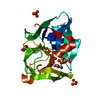
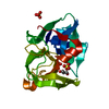
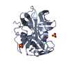
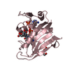
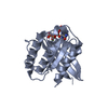
- PDB-1ssx: 0.83A resolution crystal structure of alpha-lytic protease at pH 8 -
+ Open data

- Basic information
| Entry | Database: PDB / ID: 1ssx | ||||||
|---|---|---|---|---|---|---|---|
| Title | 0.83A resolution crystal structure of alpha-lytic protease at pH 8 | ||||||
 Components Components | Alpha-lytic protease | ||||||
 Keywords Keywords | HYDROLASE / a-lytic protease / serine protease / protein folding / protein stability / packing distortion | ||||||
| Function / homology |  Function and homology information Function and homology informationalpha-lytic endopeptidase / serine-type endopeptidase activity / proteolysis / extracellular region Similarity search - Function | ||||||
| Biological species |  Lysobacter enzymogenes (bacteria) Lysobacter enzymogenes (bacteria) | ||||||
| Method |  X-RAY DIFFRACTION / SYNCHROTRON / REFINEMENT OF 1TAL.PDB / Resolution: 0.83 Å X-RAY DIFFRACTION / SYNCHROTRON / REFINEMENT OF 1TAL.PDB / Resolution: 0.83 Å | ||||||
 Authors Authors | Fuhrmann, C.N. / Agard, D.A. | ||||||
 Citation Citation | Journal: J.Mol.Biol. / Year: 2004 Title: The 0.83A Resolution Crystal Structure of alpha-Lytic Protease Reveals the Detailed Structure of the Active Site and Identifies a Source of Conformational Strain. Authors: Fuhrmann, C.N. / Kelch, B.A. / Ota, N. / Agard, D.A. | ||||||
| History |
|
- Structure visualization
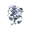
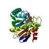
| Structure viewer | Molecule: MolmilJmol/JSmol |
|---|
- Downloads & links
-Download
| PDBx/mmCIF format | 1ssx.cif.gz | 144.9 KB | Display | PDBx/mmCIF format |
|---|---|---|---|---|
| PDB format | pdb1ssx.ent.gz | 116.7 KB | Display | PDB format |
| PDBx/mmJSON format | 1ssx.json.gz | Tree view | PDBx/mmJSON format | |
| Others |  Other downloads Other downloads |
-Validation report
| Arichive directory | https://data.pdbj.org/pub/pdb/validation_reports/ss/1ssxftp://data.pdbj.org/pub/pdb/validation_reports/ss/1ssx | HTTPS FTP |
|---|
-Related structure data
| Related structure data |  1talS S: Starting model for refinement |
|---|---|
| Similar structure data |


-Links
 PDBj
PDBj- Assembly
| Deposited unit | 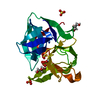
| |||||||||||||||
|---|---|---|---|---|---|---|---|---|---|---|---|---|---|---|---|---|
| 1 |
| |||||||||||||||
| Unit cell |
| |||||||||||||||
| Components on special symmetry positions |
|
-Components
| #1: Protein | Mass: 19875.131 Da / Num. of mol.: 1 / Fragment: Mature protease domain (residues 200-397) Source method: isolated from a genetically manipulated source Source: (gene. exp.) Lysobacter enzymogenes (bacteria) / Gene: ALPHA-LP / Plasmid: pALP12 / Production host: | ||||||
|---|---|---|---|---|---|---|---|
| #2: Chemical | ChemComp-SO4 /   Mass: 96.063 Da / Num. of mol.: 4 / Source method: obtained synthetically / Formula: SO4 Mass: 96.063 Da / Num. of mol.: 4 / Source method: obtained synthetically / Formula: SO4#3: Chemical |   Mass: 92.094 Da / Num. of mol.: 2 / Source method: obtained synthetically / Formula: C3H8O3 Mass: 92.094 Da / Num. of mol.: 2 / Source method: obtained synthetically / Formula: C3H8O3#4: Water | ChemComp-HOH / |  Mass: 18.015 Da / Num. of mol.: 467 / Source method: isolated from a natural source / Formula: H2O Mass: 18.015 Da / Num. of mol.: 467 / Source method: isolated from a natural source / Formula: H2OHas protein modification | Y | |
-Experimental details
-Experiment
| Experiment | Method: X-RAY DIFFRACTION / Number of used crystals: 1 |
|---|
- Sample preparation
| Crystal | Density Matthews: 2.51 Å3/Da / Density % sol: 51 % |
|---|---|
| Crystal grow | Temperature: 298 K / Method: vapor diffusion, hanging drop / pH: 8 Details: 1.3M lithium sulfate, 0.02M Tris, pH 8.0, VAPOR DIFFUSION, HANGING DROP, temperature 298K |
-Data collection
| Diffraction | Mean temperature: 100 K |
|---|---|
| Diffraction source | Source: SYNCHROTRON / Site: SSRL  / Beamline: BL9-1 / Wavelength: 0.785 Å / Beamline: BL9-1 / Wavelength: 0.785 Å |
| Detector | Type: MARRESEARCH / Detector: IMAGE PLATE / Date: Jan 7, 2000 Details: flat mirror (vertical focusing); single crystal Si(311) bent monochromator (horizontal focusing) |
| Radiation | Monochromator: single crystal Si(311) bent monochromator / Protocol: SINGLE WAVELENGTH / Monochromatic (M) / Laue (L): M / Scattering type: x-ray |
| Radiation wavelength | Wavelength: 0.785 Å / Relative weight: 1 |
| Reflection | Resolution: 0.83→10 Å / Num. all: 187431 / Num. obs: 187431 / % possible obs: 99.6 % / Redundancy: 4.3 % / Rmerge(I) obs: 0.059 / Net I/σ(I): 28.5 |
| Reflection shell | Resolution: 0.83→0.84 Å / Redundancy: 2.8 % / Rmerge(I) obs: 0.407 / Mean I/σ(I) obs: 3 / Num. unique all: 9176 / % possible all: 98.8 |
- Processing
| Software |
| |||||||||||||||||||||||||||||||||
|---|---|---|---|---|---|---|---|---|---|---|---|---|---|---|---|---|---|---|---|---|---|---|---|---|---|---|---|---|---|---|---|---|---|---|
| Refinement | Method to determine structure: REFINEMENT OF 1TAL.PDB Starting model: PDB ENTRY 1TAL Resolution: 0.83→10 Å / Num. parameters: 19125 / Num. restraintsaints: 20884 / Isotropic thermal model: anisotropic / Cross valid method: FREE R / σ(F): 0 / σ(I): 0 / Stereochemistry target values: Engh & Huber Details: Hydrogen atoms were included in the refinement as "riding hydrogens", with position and geometry fixed to those values defined by SHELXL-97. Methyl and hydroxyl hydrogens on single-conformer ...Details: Hydrogen atoms were included in the refinement as "riding hydrogens", with position and geometry fixed to those values defined by SHELXL-97. Methyl and hydroxyl hydrogens on single-conformer sidechains were each positioned at a torsion angle that best satisfied positive difference electron density (using instructions HFIX 137 and HFIX 147, respectively). It should be noted that the length of donor-hydrogen bonds in this structure are likely shorter than their true internuclear distance; these bond lengths are defined by SHELXL-97 parameters. The positions of only four hydrogen atoms were allowed to refine freely: His57 HD1, His57 HE1, Ser214 HG, and Gly193 HN. During the final stages of refinement, geometrical restraints were released for all non-hydrogen atoms in residues with single conformations. See publication for more details.
| |||||||||||||||||||||||||||||||||
| Solvent computation | Solvent model: MOEWS & KRETSINGER, J.MOL.BIOL.91(1973)201-228 | |||||||||||||||||||||||||||||||||
| Displacement parameters | Biso mean: 10.8 Å2 | |||||||||||||||||||||||||||||||||
| Refine analyze | Num. disordered residues: 23 / Occupancy sum hydrogen: 1362.61 / Occupancy sum non hydrogen: 1767.2 | |||||||||||||||||||||||||||||||||
| Refinement step | Cycle: LAST / Resolution: 0.83→10 Å
| |||||||||||||||||||||||||||||||||
| Refine LS restraints |
|
