 Movie
Movie Controller

[English] 日本語
 Yorodumi
- PDB-2alp: REFINED STRUCTURE OF ALPHA-LYTIC PROTEASE AT 1.7 ANGSTROMS RESOLU... -
+ Open data

- Basic information
| Entry | Database: PDB / ID: 2alp | |||||||||
|---|---|---|---|---|---|---|---|---|---|---|
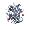
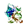
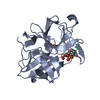
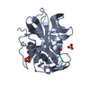
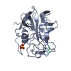
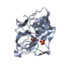
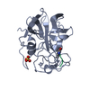
| Title | REFINED STRUCTURE OF ALPHA-LYTIC PROTEASE AT 1.7 ANGSTROMS RESOLUTION. ANALYSIS OF HYDROGEN BONDING AND SOLVENT STRUCTURE | |||||||||
 Components Components | ALPHA-LYTIC PROTEASE | |||||||||
 Keywords Keywords | HYDROLASE (SERINE PROTEINASE) | |||||||||
| Function / homology |  Function and homology information Function and homology informationalpha-lytic endopeptidase / serine-type endopeptidase activity / proteolysis / extracellular region Similarity search - Function | |||||||||
| Biological species |  Lysobacter enzymogenes (bacteria) Lysobacter enzymogenes (bacteria) | |||||||||
| Method |  X-RAY DIFFRACTION / Resolution: 1.7 Å X-RAY DIFFRACTION / Resolution: 1.7 Å | |||||||||
 Authors Authors | Fujinaga, M. / Delbaere, L.T.J. / Brayer, G.D. / James, M.N.G. | |||||||||
 Citation Citation | Journal: J.Mol.Biol. / Year: 1985 Title: Refined structure of alpha-lytic protease at 1.7 A resolution. Analysis of hydrogen bonding and solvent structure. Authors: Fujinaga, M. / Delbaere, L.T. / Brayer, G.D. / James, M.N. #1: Journal: J.Mol.Biol. / Year: 1979Title: Molecular Structure of the Alpha-Lytic Protease from Myxobacter 495 at 2.8 Angstroms Resolution Authors: Brayer, G.D. / Delbaere, L.T.J. / James, M.N.G. #2: Journal: Nature / Year: 1979Title: Comparison of the Predicted Model of Alph-Lytic Protease with the X-Ray Structure Authors: Delbaere, L.T.J. / Brayer, G.D. / James, M.N.G. #3: Journal: Can.J.Biochem. / Year: 1978Title: Amino Acid Sequence Alignment of Bacterial and Mammalian Pancreatic Serine Proteases Based on Topological Equivalences Authors: James, M.N.G. / Delbaere, L.T.J. / Brayer, G.D. | |||||||||
| History |
|
- Structure visualization
| Structure viewer | Molecule: MolmilJmol/JSmol |
|---|
- Downloads & links
-Download
| PDBx/mmCIF format | 2alp.cif.gz | 50.7 KB | Display | PDBx/mmCIF format |
|---|---|---|---|---|
| PDB format | pdb2alp.ent.gz | 35.7 KB | Display | PDB format |
| PDBx/mmJSON format | 2alp.json.gz | Tree view | PDBx/mmJSON format | |
| Others |  Other downloads Other downloads |
-Validation report
| Arichive directory | https://data.pdbj.org/pub/pdb/validation_reports/al/2alpftp://data.pdbj.org/pub/pdb/validation_reports/al/2alp | HTTPS FTP |
|---|
-Related structure data
| Similar structure data |
|---|


-Links
 PDBj
PDBj- Assembly
| Deposited unit | 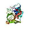
| ||||||||
|---|---|---|---|---|---|---|---|---|---|
| 1 |
| ||||||||
| Unit cell |
| ||||||||
| Atom site foot note | 1: RESIDUE 95 IS A CIS-PROLINE. |
-Components
| #1: Protein | Mass: 19875.131 Da / Num. of mol.: 1 Source method: isolated from a genetically manipulated source Source: (gene. exp.) Lysobacter enzymogenes (bacteria) / References: UniProt: P00778, alpha-lytic endopeptidase | ||||
|---|---|---|---|---|---|
| #2: Chemical |   Mass: 96.063 Da / Num. of mol.: 2 / Source method: obtained synthetically / Formula: SO4 Mass: 96.063 Da / Num. of mol.: 2 / Source method: obtained synthetically / Formula: SO4#3: Water | ChemComp-HOH / |  Mass: 18.015 Da / Num. of mol.: 156 / Source method: isolated from a natural source / Formula: H2O Mass: 18.015 Da / Num. of mol.: 156 / Source method: isolated from a natural source / Formula: H2OHas protein modification | Y | |
-Experimental details
-Experiment
| Experiment | Method: X-RAY DIFFRACTION |
|---|
- Sample preparation
| Crystal | Density Matthews: 2.55 Å3/Da / Density % sol: 51.7 % | ||||||||||||||||||
|---|---|---|---|---|---|---|---|---|---|---|---|---|---|---|---|---|---|---|---|
| Crystal grow | *PLUS pH: 6.1 / Method: microdialysis / Details: Brayer, G.D., (1979) J.Mol.Biol., 131, 743. | ||||||||||||||||||
| Components of the solutions | *PLUS
|
-Data collection
| Reflection | *PLUS |
|---|
- Processing
| Refinement | Resolution: 1.7→8 Å / Rfactor Rwork: 0.131 / σ(I): 2 | ||||||||||||||||||||||||||||||||||||||||||||||||||||||||||||
|---|---|---|---|---|---|---|---|---|---|---|---|---|---|---|---|---|---|---|---|---|---|---|---|---|---|---|---|---|---|---|---|---|---|---|---|---|---|---|---|---|---|---|---|---|---|---|---|---|---|---|---|---|---|---|---|---|---|---|---|---|---|
| Refinement step | Cycle: LAST / Resolution: 1.7→8 Å
| ||||||||||||||||||||||||||||||||||||||||||||||||||||||||||||
| Refine LS restraints |
| ||||||||||||||||||||||||||||||||||||||||||||||||||||||||||||
| Refinement | *PLUS Num. reflection obs: 14996 / σ(I): 2 / Highest resolution: 1.7 Å / Lowest resolution: 8 Å / Rfactor obs: 0.131 | ||||||||||||||||||||||||||||||||||||||||||||||||||||||||||||
| Solvent computation | *PLUS | ||||||||||||||||||||||||||||||||||||||||||||||||||||||||||||
| Displacement parameters | *PLUS Biso mean: 14.3 Å2 | ||||||||||||||||||||||||||||||||||||||||||||||||||||||||||||
| Refine LS restraints | *PLUS
|
