 Movie
Movie Controller


+ Open data

- Basic information
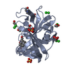
| Entry | Database: PDB / ID: 1tal | ||||||
|---|---|---|---|---|---|---|---|
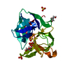
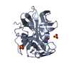
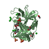
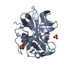
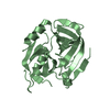
| Title | ALPHA-LYTIC PROTEASE AT 120 K (SINGLE STRUCTURE MODEL) | ||||||
 Components Components | ALPHA-LYTIC PROTEASE | ||||||
 Keywords Keywords | SERINE PROTEASE / LOW TEMPERATURE / HYDROLASE / SERINE PROTEINASE | ||||||
| Function / homology |  Function and homology information Function and homology informationalpha-lytic endopeptidase / serine-type endopeptidase activity / proteolysis / extracellular region Similarity search - Function | ||||||
| Biological species |  Lysobacter enzymogenes (bacteria) Lysobacter enzymogenes (bacteria) | ||||||
| Method |  X-RAY DIFFRACTION / SYNCHROTRON / REFINEMENT OF 2ALP / Resolution: 1.5 Å X-RAY DIFFRACTION / SYNCHROTRON / REFINEMENT OF 2ALP / Resolution: 1.5 Å | ||||||
 Authors Authors | Rader, S.D. / Agard, D.A. | ||||||
 Citation Citation | Journal: Protein Sci. / Year: 1997 Title: Conformational substates in enzyme mechanism: the 120 K structure of alpha-lytic protease at 1.5 A resolution. Authors: Rader, S.D. / Agard, D.A. #1: Journal: Biochemistry / Year: 1991Title: Structural Basis for Broad Specificity in Alpha-Lytic Protease Authors: Bone, R. / Fujishige, A. / Kettner, C.A. / Agard, D.A. #2: Journal: Nature / Year: 1989Title: Structural Plasticity Broadens the Specificity of an Engineered Protease Authors: Bone, R. / Silen, J.L. / Agard, D.A. #3: Journal: Biochemistry / Year: 1989Title: Structural Analysis of Specificity: Alpha-Lytic Protease Complexes with Analogues of Reaction Intermediates Authors: Bone, R. / Frank, D. / Kettner, C.A. / Agard, D.A. #4: Journal: Biochemistry / Year: 1987Title: Serine Protease Mechanism: Structure of an Inhibitory Complex of Alpha-Lytic Protease and a Tightly Bound Peptide Boronic Acid Authors: Bone, R. / Shenvi, A.B. / Kettner, C.A. / Agard, D.A. #5: Journal: J.Mol.Biol. / Year: 1985Title: Refined Structure of Alpha-Lytic Protease at 1.7 A Resolution. Analysis of Hydrogen Bonding and Solvent Structure Authors: Fujinaga, M. / Delbaere, L.T. / Brayer, G.D. / James, M.N. #6: Journal: J.Mol.Biol. / Year: 1979Title: Molecular Structure of the Alpha-Lytic Protease from Myxobacter 495 at 2.8 Angstroms Resolution Authors: Brayer, G.D. / Delbaere, L.T. / James, M.N. | ||||||
| History |
|
- Structure visualization
| Structure viewer | Molecule: MolmilJmol/JSmol |
|---|
- Downloads & links
-Download
| PDBx/mmCIF format | 1tal.cif.gz | 58 KB | Display | PDBx/mmCIF format |
|---|---|---|---|---|
| PDB format | pdb1tal.ent.gz | 40.9 KB | Display | PDB format |
| PDBx/mmJSON format | 1tal.json.gz | Tree view | PDBx/mmJSON format | |
| Others |  Other downloads Other downloads |
-Validation report
| Arichive directory | https://data.pdbj.org/pub/pdb/validation_reports/ta/1talftp://data.pdbj.org/pub/pdb/validation_reports/ta/1tal | HTTPS FTP |
|---|
-Related structure data


-Links
 PDBj
PDBj- Assembly
| Deposited unit | 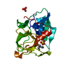
| ||||||||
|---|---|---|---|---|---|---|---|---|---|
| 1 |
| ||||||||
| Unit cell |
|
-Components
| #1: Protein | Mass: 19875.131 Da / Num. of mol.: 1 Source method: isolated from a genetically manipulated source Source: (gene. exp.) Lysobacter enzymogenes (bacteria) / Strain: 495 / Production host: | ||||||||||
|---|---|---|---|---|---|---|---|---|---|---|---|
| #2: Chemical |   Mass: 96.063 Da / Num. of mol.: 3 / Source method: obtained synthetically / Formula: SO4 Mass: 96.063 Da / Num. of mol.: 3 / Source method: obtained synthetically / Formula: SO4#3: Chemical | ChemComp-TAM / |   Mass: 163.215 Da / Num. of mol.: 1 / Source method: obtained synthetically / Formula: C7H17NO3 / Comment: pH buffer*YM Mass: 163.215 Da / Num. of mol.: 1 / Source method: obtained synthetically / Formula: C7H17NO3 / Comment: pH buffer*YM#4: Water | ChemComp-HOH / |  Mass: 18.015 Da / Num. of mol.: 301 / Source method: isolated from a natural source / Formula: H2O Mass: 18.015 Da / Num. of mol.: 301 / Source method: isolated from a natural source / Formula: H2OHas protein modification | Y | Nonpolymer details | TERMINAL OXYGENS OF THE TRIS MOLECULE ARE NOT SEEN IN THE DENSITY. | Sequence details | RESIDUE NUMBERING IS BY HOMOLOGY WITH CHYMOTRYPS | |
-Experimental details
-Experiment
| Experiment | Method: X-RAY DIFFRACTION / Number of used crystals: 1 |
|---|
- Sample preparation
| Crystal | Density Matthews: 2.5 Å3/Da / Density % sol: 48.2 % | ||||||||||||||||||||||||
|---|---|---|---|---|---|---|---|---|---|---|---|---|---|---|---|---|---|---|---|---|---|---|---|---|---|
| Crystal grow | pH: 8 / Details: pH 8.0 | ||||||||||||||||||||||||
| Crystal grow | *PLUS pH: 7.5 / Method: vapor diffusion, hanging drop | ||||||||||||||||||||||||
| Components of the solutions | *PLUS
|
-Data collection
| Diffraction | Mean temperature: 120 K |
|---|---|
| Diffraction source | Source: SYNCHROTRON / Site: SSRL  / Beamline: BL7-1 / Wavelength: 1.08 / Beamline: BL7-1 / Wavelength: 1.08 |
| Detector | Type: MARRESEARCH / Detector: IMAGE PLATE / Date: Jul 25, 1992 |
| Radiation | Monochromatic (M) / Laue (L): M / Scattering type: x-ray |
| Radiation wavelength | Wavelength: 1.08 Å / Relative weight: 1 |
| Reflection | Resolution: 1.5→20 Å / Num. obs: 33422 / % possible obs: 99 % / Observed criterion σ(I): -3 / Redundancy: 4.7 % / Biso Wilson estimate: 5.8 Å2 / Rmerge(I) obs: 0.059 |
| Reflection shell | Resolution: 1.5→1.57 Å / % possible all: 99.1 |
| Reflection | *PLUS Num. obs: 31458 / Num. measured all: 148867 |
| Reflection shell | *PLUS % possible obs: 99.1 % |
- Processing
| Software |
| ||||||||||||||||||||||||||||||||||||||||||||||||||||||||||||
|---|---|---|---|---|---|---|---|---|---|---|---|---|---|---|---|---|---|---|---|---|---|---|---|---|---|---|---|---|---|---|---|---|---|---|---|---|---|---|---|---|---|---|---|---|---|---|---|---|---|---|---|---|---|---|---|---|---|---|---|---|---|
| Refinement | Method to determine structure: REFINEMENT OF 2ALP / Resolution: 1.5→6 Å / Isotropic thermal model: UNRESTRAINED / Cross valid method: FREE R / σ(F): 0
| ||||||||||||||||||||||||||||||||||||||||||||||||||||||||||||
| Displacement parameters | Biso mean: 5.36 Å2 | ||||||||||||||||||||||||||||||||||||||||||||||||||||||||||||
| Refinement step | Cycle: LAST / Resolution: 1.5→6 Å
| ||||||||||||||||||||||||||||||||||||||||||||||||||||||||||||
| Refine LS restraints |
| ||||||||||||||||||||||||||||||||||||||||||||||||||||||||||||
| LS refinement shell | Resolution: 1.5→1.57 Å / Total num. of bins used: 8
| ||||||||||||||||||||||||||||||||||||||||||||||||||||||||||||
| Xplor file |
| ||||||||||||||||||||||||||||||||||||||||||||||||||||||||||||
| Software | *PLUS Name: X-PLOR / Classification: refinement | ||||||||||||||||||||||||||||||||||||||||||||||||||||||||||||
| Refinement | *PLUS | ||||||||||||||||||||||||||||||||||||||||||||||||||||||||||||
| Solvent computation | *PLUS | ||||||||||||||||||||||||||||||||||||||||||||||||||||||||||||
| Displacement parameters | *PLUS | ||||||||||||||||||||||||||||||||||||||||||||||||||||||||||||
| Refine LS restraints | *PLUS
| ||||||||||||||||||||||||||||||||||||||||||||||||||||||||||||
| LS refinement shell | *PLUS Rfactor obs: 0.1897 |
