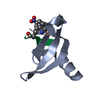
 Movie
Movie Controller


+ Open data

- Basic information
| Entry | Database: PDB / ID: 7baf | |||||||||
|---|---|---|---|---|---|---|---|---|---|---|
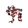
| Title | Crystal structure of PAFB in complex with zinc | |||||||||
 Components Components | Antifungal protein | |||||||||
 Keywords Keywords | ANTIFUNGAL PROTEIN / Penicillium chrysogenum antifungal protein B / calix[n]arene / zinc / porous framework / molecular recognition | |||||||||
| Function / homology | Antifungal protein / Antifungal protein domain superfamily / Antifungal protein / defense response to fungus / killing of cells of another organism / host cell cytoplasm / extracellular region / ACETATE ION / Antifungal protein B Function and homology information Function and homology information | |||||||||
| Biological species |  Penicillium rubens (fungus) Penicillium rubens (fungus) | |||||||||
| Method |  X-RAY DIFFRACTION / SYNCHROTRON / MOLECULAR REPLACEMENT / Resolution: 1.123 Å X-RAY DIFFRACTION / SYNCHROTRON / MOLECULAR REPLACEMENT / Resolution: 1.123 Å | |||||||||
 Authors Authors | Guagnini, F. / Huber, A. / Alex, J.M. / Marx, F. / Crowley, P.B. | |||||||||
| Funding support |  Ireland, 2items Ireland, 2items
| |||||||||
 Citation Citation | Journal: J.Struct.Biol. / Year: 2021 Title: Porous assembly of an antifungal protein mediated by zinc and sulfonato-calix[8]arene. Authors: Guagnini, F. / Huber, A. / Alex, J.M. / Marx, F. / Crowley, P.B. | |||||||||
| History |
|
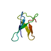
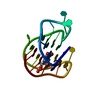
- Structure visualization
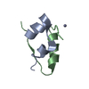
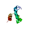
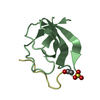
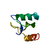
| Structure viewer | Molecule: MolmilJmol/JSmol |
|---|
- Downloads & links
-Download
| PDBx/mmCIF format | 7baf.cif.gz | 39.2 KB | Display | PDBx/mmCIF format |
|---|---|---|---|---|
| PDB format | pdb7baf.ent.gz | 25 KB | Display | PDB format |
| PDBx/mmJSON format | 7baf.json.gz | Tree view | PDBx/mmJSON format | |
| Others |  Other downloads Other downloads |
-Validation report
| Arichive directory | https://data.pdbj.org/pub/pdb/validation_reports/ba/7bafftp://data.pdbj.org/pub/pdb/validation_reports/ba/7baf | HTTPS FTP |
|---|
-Related structure data
| Related structure data | 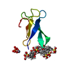 7badC  7baeC 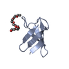 6hajS S: Starting model for refinement C: citing same article ( |
|---|---|
| Similar structure data |


-Links
 PDBj
PDBj- Assembly
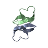
| Deposited unit | 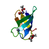
| ||||||||
|---|---|---|---|---|---|---|---|---|---|
| 1 |
| ||||||||
| Unit cell |
|
-Components
| #1: Protein | Mass: 6516.394 Da / Num. of mol.: 1 Source method: isolated from a genetically manipulated source Source: (gene. exp.) Penicillium rubens (strain ATCC 28089 / DSM 1075 / NRRL 1951 / Wisconsin 54-1255) (fungus)Strain: ATCC 28089 / DSM 1075 / NRRL 1951 / Wisconsin 54-1255 Gene: afp, Pc12g08290 Production host: Penicillium rubens Wisconsin 54-1255 (fungus)References: UniProt: B6GXZ8 | ||||||||
|---|---|---|---|---|---|---|---|---|---|
| #2: Chemical | ChemComp-ACT /   Mass: 59.044 Da / Num. of mol.: 5 / Source method: obtained synthetically / Formula: C2H3O2 Mass: 59.044 Da / Num. of mol.: 5 / Source method: obtained synthetically / Formula: C2H3O2#3: Chemical |   Mass: 65.409 Da / Num. of mol.: 3 / Source method: obtained synthetically / Formula: Zn / Feature type: SUBJECT OF INVESTIGATION Mass: 65.409 Da / Num. of mol.: 3 / Source method: obtained synthetically / Formula: Zn / Feature type: SUBJECT OF INVESTIGATION#4: Water | ChemComp-HOH / |  Mass: 18.015 Da / Num. of mol.: 48 / Source method: isolated from a natural source / Formula: H2O Mass: 18.015 Da / Num. of mol.: 48 / Source method: isolated from a natural source / Formula: H2OHas ligand of interest | Y | Has protein modification | Y | |
-Experimental details
-Experiment
| Experiment | Method: X-RAY DIFFRACTION / Number of used crystals: 1 |
|---|
- Sample preparation
| Crystal | Density Matthews: 2.06 Å3/Da / Density % sol: 40 % |
|---|---|
| Crystal grow | Temperature: 293.15 K / Method: vapor diffusion, sitting drop / pH: 5.5 Details: 0.1 M BIS-TRIS pH 5.5, 25 % PEG 3350, 0.2 M NH4CH3COO |
-Data collection
| Diffraction | Mean temperature: 100 K / Serial crystal experiment: N | ||||||||||||||||||||||||||||||
|---|---|---|---|---|---|---|---|---|---|---|---|---|---|---|---|---|---|---|---|---|---|---|---|---|---|---|---|---|---|---|---|
| Diffraction source | Source: SYNCHROTRON / Site: SOLEIL  / Beamline: PROXIMA 2 / Wavelength: 0.98013 Å / Beamline: PROXIMA 2 / Wavelength: 0.98013 Å | ||||||||||||||||||||||||||||||
| Detector | Type: DECTRIS EIGER2 X 9M / Detector: PIXEL / Date: Sep 21, 2019 | ||||||||||||||||||||||||||||||
| Radiation | Protocol: SINGLE WAVELENGTH / Monochromatic (M) / Laue (L): M / Scattering type: x-ray | ||||||||||||||||||||||||||||||
| Radiation wavelength | Wavelength: 0.98013 Å / Relative weight: 1 | ||||||||||||||||||||||||||||||
| Reflection | Resolution: 1.12→28.53 Å / Num. obs: 19859 / % possible obs: 99.9 % / Redundancy: 9.4 % / Biso Wilson estimate: 7.96 Å2 / CC1/2: 0.995 / Rmerge(I) obs: 0.163 / Rpim(I) all: 0.055 / Rrim(I) all: 0.172 / Net I/σ(I): 10.4 / Num. measured all: 186816 / Scaling rejects: 1456 | ||||||||||||||||||||||||||||||
| Reflection shell | Diffraction-ID: 1
|
- Processing
| Software |
| ||||||||||||||||||||||||||||||||||||||||||||||||||||||||||||||||||||||||||||||||||||||||||||||||||||||||||||
|---|---|---|---|---|---|---|---|---|---|---|---|---|---|---|---|---|---|---|---|---|---|---|---|---|---|---|---|---|---|---|---|---|---|---|---|---|---|---|---|---|---|---|---|---|---|---|---|---|---|---|---|---|---|---|---|---|---|---|---|---|---|---|---|---|---|---|---|---|---|---|---|---|---|---|---|---|---|---|---|---|---|---|---|---|---|---|---|---|---|---|---|---|---|---|---|---|---|---|---|---|---|---|---|---|---|---|---|---|---|
| Refinement | Method to determine structure: MOLECULAR REPLACEMENT Starting model: 6HAJ Resolution: 1.123→28.53 Å / Cor.coef. Fo:Fc: 0.937 / Cor.coef. Fo:Fc free: 0.953 / SU R Cruickshank DPI: 0.035 / Cross valid method: THROUGHOUT / σ(F): 0 / SU R Blow DPI: 0.035 / SU Rfree Blow DPI: 0.034 / SU Rfree Cruickshank DPI: 0.033 Details: HYDROGENS WERE FULLY REFINED WITH ZERO OCCUPANCY AT NUCLEAR POSITION. REFINEMENT NOTES. NUMBER OF REFINEMENT NOTES : 1 NOTE 1 : IDEAL-DIST CONTACT TERM CONTACT SETUP. ALL ATOMS HAVE CCP4 ATOM TYPE FROM LIBRARY
| ||||||||||||||||||||||||||||||||||||||||||||||||||||||||||||||||||||||||||||||||||||||||||||||||||||||||||||
| Displacement parameters | Biso max: 37.1 Å2 / Biso mean: 13.04 Å2 / Biso min: 5.21 Å2
| ||||||||||||||||||||||||||||||||||||||||||||||||||||||||||||||||||||||||||||||||||||||||||||||||||||||||||||
| Refine analyze | Luzzati coordinate error obs: 0.14 Å | ||||||||||||||||||||||||||||||||||||||||||||||||||||||||||||||||||||||||||||||||||||||||||||||||||||||||||||
| Refinement step | Cycle: final / Resolution: 1.123→28.53 Å
| ||||||||||||||||||||||||||||||||||||||||||||||||||||||||||||||||||||||||||||||||||||||||||||||||||||||||||||
| Refine LS restraints |
| ||||||||||||||||||||||||||||||||||||||||||||||||||||||||||||||||||||||||||||||||||||||||||||||||||||||||||||
| LS refinement shell | Resolution: 1.123→1.13 Å / Rfactor Rfree error: 0
| ||||||||||||||||||||||||||||||||||||||||||||||||||||||||||||||||||||||||||||||||||||||||||||||||||||||||||||
| Refinement TLS params. | Method: refined / Origin x: -1.713 Å / Origin y: 24.3568 Å / Origin z: 11.5421 Å
| ||||||||||||||||||||||||||||||||||||||||||||||||||||||||||||||||||||||||||||||||||||||||||||||||||||||||||||
| Refinement TLS group | Selection details: { A|* } |
