 Movie
Movie Controller


+ Open data

- Basic information
| Entry | Database: PDB / ID: 2ins | ||||||
|---|---|---|---|---|---|---|---|
| Title | THE STRUCTURE OF DES-PHE B1 BOVINE INSULIN | ||||||
 Components Components |
| ||||||
 Keywords Keywords | HORMONE | ||||||
| Function / homology |  Function and homology information Function and homology informationestradiol secretion / positive regulation of blood circulation / negative regulation of lactation / glucose import in response to insulin stimulus / positive regulation of cell maturation / response to L-arginine / positive regulation of mammary gland epithelial cell proliferation / positive regulation of lactation / response to butyrate / negative regulation of appetite ...estradiol secretion / positive regulation of blood circulation / negative regulation of lactation / glucose import in response to insulin stimulus / positive regulation of cell maturation / response to L-arginine / positive regulation of mammary gland epithelial cell proliferation / positive regulation of lactation / response to butyrate / negative regulation of appetite / feeding behavior / response to growth hormone / positive regulation of peptide hormone secretion / response to food / positive regulation of Rho protein signal transduction / protein secretion / negative regulation of lipid catabolic process / response to glucose / insulin receptor binding / positive regulation of protein secretion / hormone activity / response to nutrient levels / glucose metabolic process / positive regulation of insulin secretion / glucose homeostasis / response to heat / positive regulation of phosphatidylinositol 3-kinase/protein kinase B signal transduction / positive regulation of gene expression / negative regulation of apoptotic process / : / identical protein binding Similarity search - Function | ||||||
| Biological species |  | ||||||
| Method |  X-RAY DIFFRACTION / Resolution: 2.5 Å X-RAY DIFFRACTION / Resolution: 2.5 Å | ||||||
 Authors Authors | Smith, G.D. / Duax, W.L. / Dodson, E.J. / Dodson, G.G. / Degraaf, R.A.G. / Reynolds, C.D. | ||||||
 Citation Citation | Journal: Acta Crystallogr.,Sect.B / Year: 1982 Title: The Structure of Des-Phe B1 Bovine Insulin Authors: Smith, G.D. / Duax, W.L. / Dodson, E.J. / Dodson, G.G. / Degraaf, R.A.G. / Reynolds, C.D. #1: Journal: Proc.R.Soc.London,Ser.B / Year: 1983Title: A Comparative Assessment of the Zinc-Protein Coordination in 2Zn-Insulin as Determined by X-Ray Absorption Fine Structure (Exafs) and X-Ray Crystallography Authors: Bordas, J. / Dodson, G.G. / Grewe, H. / Koch, M.H.J. / Krebs, B. / Randall, J. #2: Journal: Can.J.Biochem. / Year: 1979Title: Structural Relationships in the Two-Zinc Insulin Hexamer Authors: Dodson, E.J. / Dodson, G.G. / Hodgkin, D.C. / Reynolds, C.D. #3: Journal: Acta Crystallogr.,Sect.A / Year: 1978Title: Experience with Fast Fourier Least Squares in the Refinement of the Crystal Structure of Rhombohedral 2-Zinc Insulin at 1.5 Angstroms Resolution Authors: Isaacs, N.W. / Agarwal, R.C. #4: Journal: J.Mol.Biol. / Year: 1978Title: Rhombohedral Insulin Crystal Transformation Authors: Bentley, G. / Dodson, G. / Lewitova, A. #5: Journal: Acta Crystallogr.,Sect.A / Year: 1976Title: A Method for Fitting Satisfactory Models to Sets of Atomic Positions in Protein Structure Refinements Authors: Dodson, E.J. / Isaacs, N.W. / Rollett, J.S. #8: Journal: Adv.Protein Chem. / Year: 1972Title: Insulin. The Structure in the Crystal and its Reflection in Chemistry and Biology Authors: Blundell, T. / Dodson, G. / Hodgkin, D. / Mercola, D. #9: Journal: Cold Spring Harbor Symp.Quant.Biol. / Year: 1972Title: The Crystal Structure of Rhombohedral 2 Zinc Insulin Authors: Blundell, T.L. / Cutfield, J.F. / Dodson, E.J. / Dodson, G.G. / Hodgkin, D.C. / Mercola, D.A. #10: Journal: Nature / Year: 1971Title: Atomic Positions in Rhombohedral 2-Zinc Insulin Crystals Authors: Blundell, T.L. / Cutfield, J.F. / Cutfield, S.M. / Dodson, E.J. / Dodson, G.G. / Hodgkin, D.C. / Mercola, D.A. / Vijayan, M. #11: Journal: Recent Prog.Horm.Res. / Year: 1971Title: X-Ray Analysis and the Structure of Insulin Authors: Blundell, T.L. / Dodson, G.G. / Dodson, E. / Hodgkin, D.C. / Vijayan, M. #12: Journal: J.Mol.Biol. / Year: 1970Title: X-Ray Diffraction Data on Some Crystalline Varieties of Insulin Authors: Baker, E.N. / Dodson, G. #13: Journal: Nature / Year: 1969Title: Structure of Rhombohedral 2 Zinc Insulin Crystals Authors: Adams, M.J. / Blundell, T.L. / Dodson, E.J. / Dodson, G.G. / Vijayan, M. / Baker, E.N. / Harding, M.M. / Hodgkin, D.C. / Rimmer, B. / Sheat, S. | ||||||
| History |
|
- Structure visualization
| Structure viewer | Molecule: MolmilJmol/JSmol |
|---|
- Downloads & links
-Download
| PDBx/mmCIF format | 2ins.cif.gz | 39.7 KB | Display | PDBx/mmCIF format |
|---|---|---|---|---|
| PDB format | pdb2ins.ent.gz | 27 KB | Display | PDB format |
| PDBx/mmJSON format | 2ins.json.gz | Tree view | PDBx/mmJSON format | |
| Others |  Other downloads Other downloads |
-Validation report
| Arichive directory | https://data.pdbj.org/pub/pdb/validation_reports/in/2insftp://data.pdbj.org/pub/pdb/validation_reports/in/2ins | HTTPS FTP |
|---|
-Related structure data
| Similar structure data |
|---|
-Links
 PDBj
PDBj


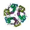
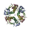
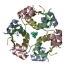
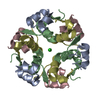
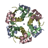
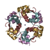
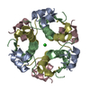
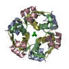
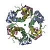
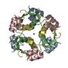
- Assembly
| Deposited unit | 
| |||||||||||||||
|---|---|---|---|---|---|---|---|---|---|---|---|---|---|---|---|---|
| 1 | 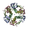
| |||||||||||||||
| 2 | 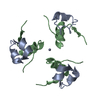
| |||||||||||||||
| 3 | 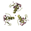
| |||||||||||||||
| 4 | 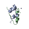
| |||||||||||||||
| 5 | 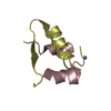
| |||||||||||||||
| Unit cell |
| |||||||||||||||
| Atom site foot note | 1: THE QUASI-TWO-FOLD SYMMETRY BREAKS DOWN MOST SERIOUSLY AT RESIDUES GLY A 1 TO GLN A 5 AND GLY C 1 TO GLN C 5 HIS B 5 AND HIS D 5 PHE B 25 AND PHE D 25 2: THE FOLLOWING RESIDUES ARE DISORDERED - ARG B 22, LYS D 29. 3: SEE REMARK 8. | |||||||||||||||
| Components on special symmetry positions |
| |||||||||||||||
| Noncrystallographic symmetry (NCS) | NCS oper: (Code: given Matrix: (-0.88, -0.48, 0.02), Details | THE CRYSTALLOGRAPHIC ASYMMETRIC UNIT OF INSULIN CONSISTS OF TWO INSULIN MOLECULES EACH CONSISTING OF TWO CHAINS. THIS ENTRY PRESENTS COORDINATES FOR MOLECULES I (CHAIN INDICATORS A AND B) AND II (CHAIN INDICATORS C AND D). THE QUASI-TWO-FOLD AXIS THAT TRANSFORMS MOLECULE I INTO MOLECULE II IS GIVEN IN THE MTRIX RECORDS BELOW. APPLYING THE THREE-FOLD CRYSTALLOGRAPHIC AXIS YIELDS A HEXAMER AROUND THE AXIS. THERE ARE TWO ZINC IONS SITUATED ON THIS THREE-FOLD AXIS. COORDINATES FOR THE ZINC IONS AND SOME WATER MOLECULES ARE INCLUDED BELOW WITH A BLANK CHAIN INDICATOR. | |
-Components
| #1: Protein/peptide | Mass: 2339.645 Da / Num. of mol.: 2 Source method: isolated from a genetically manipulated source Source: (gene. exp.) #2: Protein/peptide | Mass: 3256.753 Da / Num. of mol.: 2 Source method: isolated from a genetically manipulated source Source: (gene. exp.) #3: Chemical |   Mass: 65.409 Da / Num. of mol.: 2 / Source method: obtained synthetically / Formula: Zn Mass: 65.409 Da / Num. of mol.: 2 / Source method: obtained synthetically / Formula: Zn#4: Water | ChemComp-HOH / |  Mass: 18.015 Da / Num. of mol.: 184 / Source method: isolated from a natural source / Formula: H2O Mass: 18.015 Da / Num. of mol.: 184 / Source method: isolated from a natural source / Formula: H2OHas protein modification | Y | |
|---|
-Experimental details
-Experiment
| Experiment | Method: X-RAY DIFFRACTION |
|---|
- Sample preparation
| Crystal | Density Matthews: 1.95 Å3/Da / Density % sol: 36.8 % | ||||||||||||||||||||||||||||||||||||||||||
|---|---|---|---|---|---|---|---|---|---|---|---|---|---|---|---|---|---|---|---|---|---|---|---|---|---|---|---|---|---|---|---|---|---|---|---|---|---|---|---|---|---|---|---|
| Crystal grow | *PLUS pH: 6.5 / Method: batch method | ||||||||||||||||||||||||||||||||||||||||||
| Components of the solutions | *PLUS
|
-Data collection
| Radiation | Scattering type: x-ray |
|---|---|
| Radiation wavelength | Relative weight: 1 |
| Reflection | *PLUS Highest resolution: 2.5 Å / Num. obs: 2722 |
- Processing
| Software |
| ||||||||||||||||||||||||||||||||||||||||||||||||||||||||||||
|---|---|---|---|---|---|---|---|---|---|---|---|---|---|---|---|---|---|---|---|---|---|---|---|---|---|---|---|---|---|---|---|---|---|---|---|---|---|---|---|---|---|---|---|---|---|---|---|---|---|---|---|---|---|---|---|---|---|---|---|---|---|
| Refinement | Resolution: 2.5→5.945 Å Details: THE FOLLOWING RESIDUES ARE DISORDERED - ARG B 22, LYS D 29. THE MODEL OF THE WATER STRUCTURE OBTAINED FROM THE REFINEMENT OF 2 ZN PORCINE INSULIN AT 1.5 ANGSTROMS RESOLUTION WAS USED ...Details: THE FOLLOWING RESIDUES ARE DISORDERED - ARG B 22, LYS D 29. THE MODEL OF THE WATER STRUCTURE OBTAINED FROM THE REFINEMENT OF 2 ZN PORCINE INSULIN AT 1.5 ANGSTROMS RESOLUTION WAS USED THROUGHOUT THE DES-PHE B1 INSULIN REFINEMENT.
| ||||||||||||||||||||||||||||||||||||||||||||||||||||||||||||
| Refinement step | Cycle: LAST / Resolution: 2.5→5.945 Å
| ||||||||||||||||||||||||||||||||||||||||||||||||||||||||||||
| Refine LS restraints |
| ||||||||||||||||||||||||||||||||||||||||||||||||||||||||||||
| Refinement | *PLUS Rfactor obs: 0.18 | ||||||||||||||||||||||||||||||||||||||||||||||||||||||||||||
| Solvent computation | *PLUS | ||||||||||||||||||||||||||||||||||||||||||||||||||||||||||||
| Displacement parameters | *PLUS | ||||||||||||||||||||||||||||||||||||||||||||||||||||||||||||
| Refine LS restraints | *PLUS Type: o_bond_d / Dev ideal: 0.02 |
