 Movie
Movie Controller

[English] 日本語
 Yorodumi
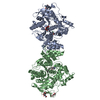
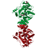
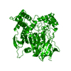
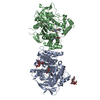
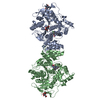
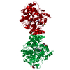
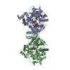
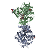
- PDB-7aiu: Crystal structure of Torpedo Californica acetylcholinesterase in ... -
+ Open data

- Basic information
| Entry | Database: PDB / ID: 7aiu | ||||||
|---|---|---|---|---|---|---|---|
| Title | Crystal structure of Torpedo Californica acetylcholinesterase in complex with 8-[(3-Chloro-6,7,10,11-tetrahydro-9-methyl-7,11-methanocycloocta[b]quinolin-12-yl)amino]-N-(4-hydroxy-3-methoxybenzyl)octanamide | ||||||
 Components Components | Acetylcholinesterase | ||||||
 Keywords Keywords | HYDROLASE / Torpedo Californica acetylcholinesterase / AD / alzheimer disease | ||||||
| Function / homology |  Function and homology information Function and homology informationacetylcholine catabolic process in synaptic cleft / choline metabolic process / acetylcholinesterase / acetylcholinesterase activity / synaptic cleft / side of membrane / synapse / extracellular space / plasma membrane Similarity search - Function | ||||||
| Biological species |   Tetronarce californica (Pacific electric ray) Tetronarce californica (Pacific electric ray) | ||||||
| Method |  X-RAY DIFFRACTION / SYNCHROTRON / MOLECULAR REPLACEMENT / Resolution: 1.996 Å X-RAY DIFFRACTION / SYNCHROTRON / MOLECULAR REPLACEMENT / Resolution: 1.996 Å | ||||||
 Authors Authors | Coquelle, N. / Colletier, J.P. | ||||||
 Citation Citation | Journal: J.Med.Chem. / Year: 2021 Title: Discovery of a Potent Dual Inhibitor of Acetylcholinesterase and Butyrylcholinesterase with Antioxidant Activity that Alleviates Alzheimer-like Pathology in Old APP/PS1 Mice. Authors: Viayna, E. / Coquelle, N. / Cieslikiewicz-Bouet, M. / Cisternas, P. / Oliva, C.A. / Sanchez-Lopez, E. / Ettcheto, M. / Bartolini, M. / De Simone, A. / Ricchini, M. / Rendina, M. / Pons, M. / ...Authors: Viayna, E. / Coquelle, N. / Cieslikiewicz-Bouet, M. / Cisternas, P. / Oliva, C.A. / Sanchez-Lopez, E. / Ettcheto, M. / Bartolini, M. / De Simone, A. / Ricchini, M. / Rendina, M. / Pons, M. / Firuzi, O. / Perez, B. / Saso, L. / Andrisano, V. / Nachon, F. / Brazzolotto, X. / Garcia, M.L. / Camins, A. / Silman, I. / Jean, L. / Inestrosa, N.C. / Colletier, J.P. / Renard, P.Y. / Munoz-Torrero, D. | ||||||
| History |
|
- Structure visualization
| Structure viewer | Molecule: MolmilJmol/JSmol |
|---|
- Downloads & links
-Download
| PDBx/mmCIF format | 7aiu.cif.gz | 245.5 KB | Display | PDBx/mmCIF format |
|---|---|---|---|---|
| PDB format | pdb7aiu.ent.gz | 193.9 KB | Display | PDB format |
| PDBx/mmJSON format | 7aiu.json.gz | Tree view | PDBx/mmJSON format | |
| Others |  Other downloads Other downloads |
-Validation report
| Arichive directory | https://data.pdbj.org/pub/pdb/validation_reports/ai/7aiuftp://data.pdbj.org/pub/pdb/validation_reports/ai/7aiu | HTTPS FTP |
|---|
-Related structure data
| Related structure data |  7aisC 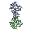 7aitC 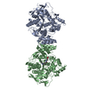 7aivC 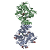 7aiwC  7aixC  7aiyC 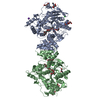 2xi4S S: Starting model for refinement C: citing same article ( |
|---|---|
| Similar structure data |


-Links
 PDBj
PDBj
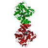
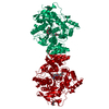
- Assembly
| Deposited unit | 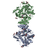
| ||||||||
|---|---|---|---|---|---|---|---|---|---|
| 1 |
| ||||||||
| Unit cell |
|
-Components
-Protein / Sugars , 2 types, 7 molecules AB
| #1: Protein | Mass: 65970.711 Da / Num. of mol.: 2 / Source method: isolated from a natural source Source: (natural) Tetronarce californica (Pacific electric ray)References: UniProt: P04058, acetylcholinesterase #3: Sugar | ChemComp-NAG /  Type: D-saccharide, beta linking / Mass: 221.208 Da / Num. of mol.: 5 / Source method: obtained synthetically / Formula: C8H15NO6 Type: D-saccharide, beta linking / Mass: 221.208 Da / Num. of mol.: 5 / Source method: obtained synthetically / Formula: C8H15NO6 |
|---|
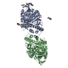
-Non-polymers , 4 types, 599 molecules 
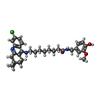


| #2: Chemical | ChemComp-PEG /  Mass: 106.120 Da / Num. of mol.: 9 / Source method: obtained synthetically / Formula: C4H10O3 Mass: 106.120 Da / Num. of mol.: 9 / Source method: obtained synthetically / Formula: C4H10O3#4: Chemical |  Mass: 564.158 Da / Num. of mol.: 2 / Source method: obtained synthetically / Formula: C33H42ClN3O3 / Feature type: SUBJECT OF INVESTIGATION Mass: 564.158 Da / Num. of mol.: 2 / Source method: obtained synthetically / Formula: C33H42ClN3O3 / Feature type: SUBJECT OF INVESTIGATION#5: Chemical |  Mass: 35.453 Da / Num. of mol.: 3 / Source method: obtained synthetically / Formula: Cl Mass: 35.453 Da / Num. of mol.: 3 / Source method: obtained synthetically / Formula: Cl#6: Water | ChemComp-HOH / | Mass: 18.015 Da / Num. of mol.: 585 / Source method: isolated from a natural source / Formula: H2O |
|---|
-Details
| Has ligand of interest | Y |
|---|---|
| Has protein modification | Y |
-Experimental details
-Experiment
| Experiment | Method: X-RAY DIFFRACTION / Number of used crystals: 1 |
|---|
- Sample preparation
| Crystal | Density Matthews: 2.77 Å3/Da / Density % sol: 55.65 % |
|---|---|
| Crystal grow | Temperature: 277 K / Method: vapor diffusion, hanging drop / Details: 28-32% PEG 200 50 mM MES / PH range: 5.8-6.2 |
-Data collection
| Diffraction | Mean temperature: 100 K / Serial crystal experiment: N |
|---|---|
| Diffraction source | Source: SYNCHROTRON / Site: ESRF  / Beamline: ID29 / Wavelength: 1 Å / Beamline: ID29 / Wavelength: 1 Å |
| Detector | Type: DECTRIS PILATUS 6M-F / Detector: PIXEL / Date: Nov 18, 2013 |
| Radiation | Protocol: SINGLE WAVELENGTH / Monochromatic (M) / Laue (L): M / Scattering type: x-ray |
| Radiation wavelength | Wavelength: 1 Å / Relative weight: 1 |
| Reflection | Resolution: 1.996→45.805 Å / Num. obs: 99278 / % possible obs: 99.1 % / Redundancy: 4.4 % / Biso Wilson estimate: 37.1 Å2 / CC1/2: 0.997 / Rmerge(I) obs: 0.094 / Net I/σ(I): 8.9 |
| Reflection shell | Resolution: 1.996→2.05 Å / Redundancy: 4.3 % / Rmerge(I) obs: 0.727 / Mean I/σ(I) obs: 1.8 / Num. unique obs: 6971 / CC1/2: 0.817 / % possible all: 95.4 |
- Processing
| Software |
| ||||||||||||||||||||||||||||||||||||||||||||||||||||||||||||||||||||||||||||||||||||||||||
|---|---|---|---|---|---|---|---|---|---|---|---|---|---|---|---|---|---|---|---|---|---|---|---|---|---|---|---|---|---|---|---|---|---|---|---|---|---|---|---|---|---|---|---|---|---|---|---|---|---|---|---|---|---|---|---|---|---|---|---|---|---|---|---|---|---|---|---|---|---|---|---|---|---|---|---|---|---|---|---|---|---|---|---|---|---|---|---|---|---|---|---|
| Refinement | Method to determine structure: MOLECULAR REPLACEMENT Starting model: 2xi4 Resolution: 1.996→45.805 Å / SU ML: 0.26 / Cross valid method: THROUGHOUT / σ(F): 1.35 / Phase error: 29.78 / Stereochemistry target values: ML
| ||||||||||||||||||||||||||||||||||||||||||||||||||||||||||||||||||||||||||||||||||||||||||
| Solvent computation | Shrinkage radii: 0.9 Å / VDW probe radii: 1.11 Å / Solvent model: FLAT BULK SOLVENT MODEL | ||||||||||||||||||||||||||||||||||||||||||||||||||||||||||||||||||||||||||||||||||||||||||
| Displacement parameters | Biso max: 120.06 Å2 / Biso mean: 36.6537 Å2 / Biso min: 19.09 Å2 | ||||||||||||||||||||||||||||||||||||||||||||||||||||||||||||||||||||||||||||||||||||||||||
| Refinement step | Cycle: final / Resolution: 1.996→45.805 Å
| ||||||||||||||||||||||||||||||||||||||||||||||||||||||||||||||||||||||||||||||||||||||||||
| Refine LS restraints |
| ||||||||||||||||||||||||||||||||||||||||||||||||||||||||||||||||||||||||||||||||||||||||||
| LS refinement shell | Refine-ID: X-RAY DIFFRACTION / Rfactor Rfree error: 0
|
